WHAT WE OFFER
Simplify your cytokine profiling studies with end-to-end support—we take care of the entire antibody array workflow, from sample preparation to final results.
Sample Processing
Send your cell lysates or protein extracts to us, and our experienced technicians will take care of all sample preparation steps according to Full Moon BioSystems’ validated protocols, ensuring consistency and compatibility with the antibody array platform.
Assay Execution
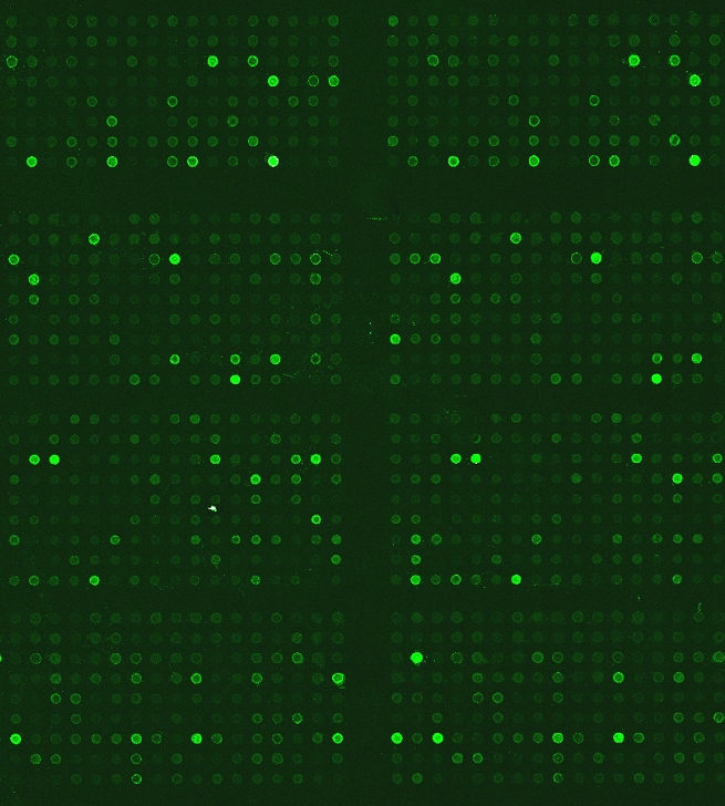
We perform the complete lysate preparation, labeling, incubation, and detection process using the Cytokine Profiling Antibody Array. This array enables high-throughput profiling of total proteins involved in key cellular signaling pathways.
Imaging & Data Analysis
Our lab uses high-resolution microarray scanners and advance software tools to extract and analyze fluorescent signals from array images. Each spot is carefully examined to verify signal quality, consistency, and accuracy across the array.
Our hybrid approach, computational quantification plus expert-reviewed, ensures high-confidence, reproducible results. We focus on delivering clean, reliable data that reflects true biological differences. This rigorous quality control process ensures that only high-confidence data are carried forward for interpretation.
Final Report Delivery
Upon completion of the assay and analysis, results are delivered in Excel format with clearly structured data. Each report includes:
-
Mean signal intensity of all replicate spots for each antibody
-
Coefficient of variation (CV) for replicate spots to assess signal consistency
-
Normalized signal intensities
-
Fold change calculations between control and. treatment samples
- Sample Assay Results
-
Biomarker profiling
-
Cancer and drug target discovery
-
Signal transduction research
-
Kinase activity studies
-
Mechanism of action studies
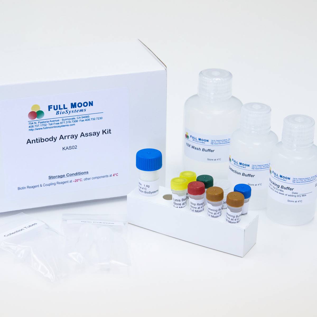
The ELISA based Phospho Explorer Antibody Array platform involves four major steps:
- Protein extraction with non-denaturing lysis buffer
- Biotinylation of protein samples
- Incubation of labeled samples with antibody array
- Detection by dye conjugated streptavidin

Cytokine Profiling Array features 310 antibodies, ideal for profiling inflammatory responses, immune regulation, and disease-related signaling.
Antibody Reactivity: Human
Selected targets :
4-1BBL, 4E-BP1, Adipolean Variant, Adiponectin, AFP, AITRL, ALCAM, Androgen receptor, Angiopoietin-1, Angiopoietin-2, APC, ApoE3, Apolipoprotein F (APOF), Apolipoprotein L1 (APOL1), Apolipoprotein L2 (APOL2), APRIL, Artemin, AXL, BAFF, BCA-1, BCL-10, BD-1, BD-2, BD-3, BD-4, BDNF, Betacellulin, BLK, BMP-2, BMP-4, BMP-7/OP-1, BRAK, Cadherin-pan, Cardiotrophin-1, Catenin-beta 1, Catenin-a1, Catenin-γ, CD14, CD40 , CD44, CEA, CIB1, C-Kit, CNTF, CTACK, CTGF, CTGFL/WISP-2, CXCL16, E-cadherin, EGF, EGFR, EGR1, EG-VEGF, EMAP-II, ENA-78, Endostatin, Eotaxin, and more.
Bae YU, You JH, Association of Protein Z with Prediabetes and Type 2 Diabetes, Endocrinol Metab (Seoul). 2021 Jun 2. doi: 10.3803/EnM.2021.962
Heo SK, Noh EK, The soluble VCAM-1 level is a potential biomarker predicting severe acute graft versus host disease after allogeneic hematopoietic cell transplantation, BMC Cancer. 2022; 22: 997
Hwang HJ, Jung SH, Identification of novel therapeutic targets in the secretome of ionizing radiation‑induced senescent tumor cells, Oncol Rep, 2016 35(2):841-850
Kang SH, Oh SY. Differential effect of cancer-associated fibroblast-derived extracellular vesicles on cisplatin resistance in oral squamous cell carcinoma via miR-876-3p. Theranostics. 2024 Jan 1;14(2):460-479. doi: 10.7150/thno.87329
Luo MXM, Wong SH, Autophagy Mediates HBx-Induced Nuclear Factor-κB Activation and Release of IL-6, IL-8 and CXCL2 in Hepatocytes, Journal of Cellular Physiology, 2015 Oct;230(10):2382-9
Shin DH, Jo JY, Midkine Is a Potential Therapeutic Target of Tumorigenesis, Angiogenesis, and Metastasis in Non-Small Cell Lung Cancer, Cancer (Basel). 2020 Aug 24;12(9):E2402. doi: 10.3390/cancers12092402
Shin E, Kim D, LDR-adapted liver-derived cytokines have potential to induce atherosclerosis, Int J Radiat Biol. 2022. DOI: 10.1080/09553002.2023.2145028
Kim DH, Cho HJ, Transplantation of PSA-NCAM-Positive Neural Precursors from Human Embryonic Stem Cells Promotes Functional Recovery in an Animal Model of Spinal Cord Injury, Tissue Eng Regen Med. 2022 Aug 29. doi: 10.1007/s13770-022-00483-z
- Cell and tissues
- Cells: > 5 million cells
- Tissues: > 75 mg
- Lysates or protein extracts
- Protein concentration: > 2 mg/mL recommended
- Protein amount: > 400 ug
- Compatible lysis buffer: mild, non-denaturing lysis, e.g., RIPA, T-per, M-per (avoid high salt/detergent concentrations)
- Serum/plasma: 20 uL
- Culture medium: protein concentration dependent
- Samples are stored at −80°C prior to shipping
- Assay Service Guide (Includes detailed sample preparation and submission instructions and forms)
- Samples must be frozen and shipped on dry ice in an insulated container
- Include sufficient dry ice for 48+ hours of transit time. Arrange shipment to ship early in the week (Monday–Wednesday for domestic shipments; Monday for international shipments) via overnight or international priority service.
- Email package tracking number to moc.oibnoomlluf@troppus to notify us of incoming shipments.
- Documentation
- A complete Sample Submission Form (MS WORD) placed inside your shipping container.
-
If submitting from outside the U.S., include required customs documents:
- Commercial invoice
-
Shipper’s Declaration stating “Non-hazardous biological sample for research use only”. This declaration must be completed on your institution’s letterhead.
Price: $1,780/sample
Note: The price listed is per sample per condition. Submitting both a control and a treatment sample counts as two samples.
U.S. Customers
-
Online Orders: credit card only
-
Purchase Orders (POs): We accept institutional or corporate POs. Email your PO to moc.oibnoomlluf@sredro.
International Customers
-
Prepayment Required: Payment must be made in advance by credit card or wire transfer.
-
Please contact us for a formal quote, invoice, and payment instructions.
-
We’ll guide you through the sample shipping process and required documentation.