Signaling Explorer Antibody Array
The Signaling Explorer Antibody Array is a high-throughput ELISA based antibody array for qualitative/semi-quantitative protein expression profiling. Featuring 1358 antibodies, this array is designed for broad screening of protein level changes in multiple cell signaling pathways, comparing normal samples to treated or diseased samples, and identifying candidate biomarkers.
Key Features
- Qualitative/semi-quantitative protein expression profiling
- Suitable samples: cell lysates; frozen or FFPE tissue lysates; serum; culture media
- Glass based array with high specificity and low background
- Antibodies are covalently attached to 3D polymer coated glass slides
- Sensitive fluorescent detection
Specifications
| Product Size: | 2 array slides per package for analyzing two samples (control vs. treated) |
| Number of Antibodies: | 1358 antibodies; 2 replicates per antibody |
| Target List: | Antibody List |
| Reactivity: | Human |
| Internal Controls: | beta-actin; GAPDH; Negative controls |
| Detection Method: | Fluorescence |Compatible Scanners |
| Slide dimensions: | 76 x 25 x 1 mm |
| Storage Condition: | 4°C for 6 months |
Product Details
14-3-3 beta, 14-3-3 epsilon, 14-3-3 eta, 14-3-3 gamma, 14-3-3 theta, 14-3-3 zeta, 4E-BP1, 5-HT-1A, 5-HT-1F, 5-HT-2C, 5-HT-3A, 5-HT-4, 5-HT-5A, A1BG, A26C2/3, AARSD1, AASDHPPT, AATF, ABCA8, ABCB7, ABCD1, ABHD11, ABHD12, ABHD12B, ABHD14A, ABHD14B, ABHD4, ABL1, ACAD10, ACBD6, ACOT2, ACOT4, ACSL6, Actin-alpha-1, Actin-gamma2, Actin-pan, ACTL6A, ACTN 1/2/3/4, ACTN alpha-2/3, ACTR-1C, ACTR3, ACVL1, ADA2L, ADAM 17, ADAR1, ADCK1, ADCK2, ADCK3, ADCK5, ADCY4, ADCY5/6, ADCY7, ADCY8, ADD2, ADD3, ADH7, ADK, ADNP, ADPGK, ADRB1, Adrenergic Receptor alpha-2A, Adrenergic Receptor alpha-2B, Adrenergic Receptor alpha-2C, Aggrecan, AGR3, and more
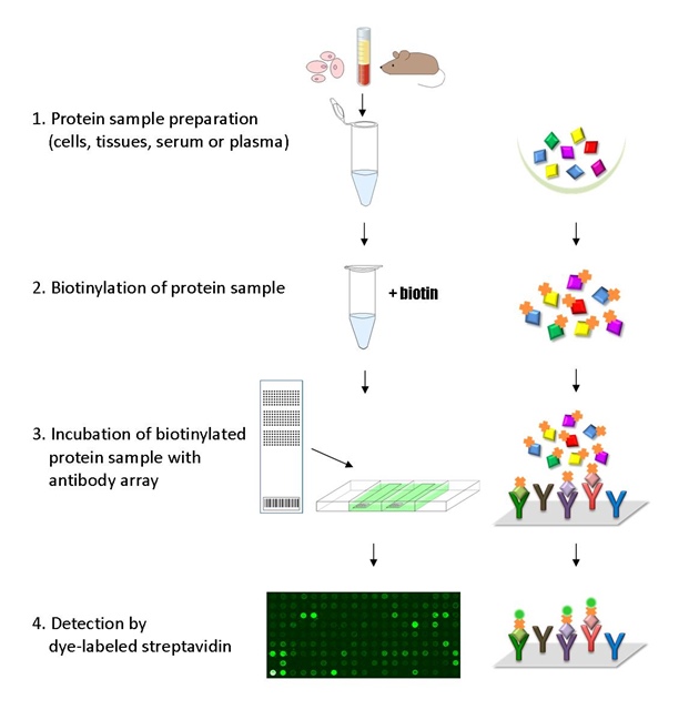
The ELISA based Cell Signaling Explorer Antibody Array platform involves four major steps: 1) Protein extraction with non-denaturing lysis buffer; 2) Biotinylation of protein samples; 3) Incubation of labeled samples with antibody array; and 4) Detection by dye conjugated streptavidin.
GAL File (To download, right click on the file name, then choose “Save target as”)
Anita L, Yin GN, Pericyte-derived extracellular vesicle-mimetic nanovesicles ameliorate erectile dysfunction via lipocalin 2 in diabetic mice, Int J Biol Sci 2022; 18(9):3653-3667. doi:10.7150/ijbs.72243
Garnett D, Caveolae as a target to quench autoinduction of the metastatic phenotype in lung cancer, J Cancer Res Clin Oncol 2016 142:611–618
Kim HJ, Choi EW, Non-thermal plasma promotes hair growth by improving the inter-follicular macroenvironment. RSC Adv. 2021. 11, 27880-27896
Klimushina MV, Gumanova NG, Direct labeling of serum proteins by fluorescent dye for antibody microarray, Biochem Biophys Res Commun 2017; 486(3):824-826
Lee J, Cho K, The Different Immune Responses by Age Are due to the Ability of the Fetal Immune System to Secrete Primal Immunoglobulins Responding to Unexperienced Antigens. Intl J. Biol Sci. 2022; 18(2): 617-636. doi: 10.7150/ijbs.67203
Lee J, Chung MY, Anacardic Acid Suppresses Adipogenesis Through Inhibition of the Hsp90/Akt Signaling Pathway in 3T3-L1 Preadipocytes, J Med Food 2021 24:5, 487-496
Lin J, Zuo J, Characterizing the molecular mechanisms of acquired temozolomide resistance in the U251 glioblastoma cell line by protein microarray, Oncol Rep, 2018 May 1;39(5):2333-41
Lund R, Leth-Larsen R, NADH-cytochrome b5 reductase 3 promotes colonization and metastasis formation and is a prognostic marker of disease-free and overall survival in estrogen receptor-negative breast cancer, Mol Cell Proteomics 2015 14(11):2988-99
Park HJ, Oh JS, Proton Irradiation Sensitizes Radioresistant Non-small Cell Lung Cancer Cells by Modulating Epidermal Growth Factor Receptor-mediated DNA Repair, Anticancer Res 2016 36(1):205-12
Rafi S, Goering J, Anti-epileptic drug topiramate upregulates TGFβ1 and SOX9 expression in primary embryonic palatal mesenchyme cells: Implications for teratogenicity, 2021 PLoS ONE 16(2): e0246989
Sakumoto M, Takahashi M, Establishment and proteomic characterization of NCC-LMS1-C1, a novel cell line of primary leiomyosarcoma of the bone, Japanese J Clin Oncol, 2017 Jul 13:1-8
Wu J, Qian J, Abstract 5547: Exploration of selective small molecule compounds as caspase inhibitors for treatment of oral mucositis caused by cancer therapy, [abstract]. In: Proceedings of the 104th Annual Meeting of the American Association for Cancer Research; 2013 Apr 6-10; Washington, DC. Philadelphia (PA): AACR; Cancer Res 2013 73(8 Suppl):Abstract nr” 5547
Zhang LL, Kwak H, An OMICS-based study of the role of C3dg in keratinocytes: RNA sequencing, antibody-chip array, and bioinformatics approaches, Int J Biol Macromol, 2019 Apr 8. pii: S0141-8130(19)31885-9
Zhao Y, Zhu W, Vascular endothelium deploys caveolin-1 to regulate oligodendrogenesis after chronic cerebral ischemia in mice, Nat Commun. 2022 Nov 10;13(1):6813. doi: 10.1038/s41467-022-34293-7
Services
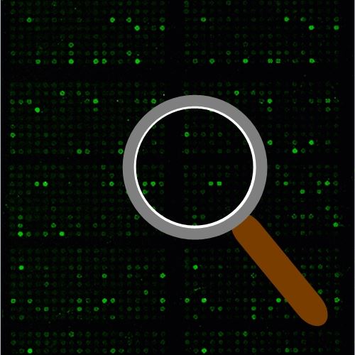
If you don’t have access to a microarray, send the finished arrays to our lab for scanning. Raw scan images are delivered in tiff format.
Cost: Free
Array Image Quantification and Analysis Service includes data extraction, data organization and analysis of the array images obtained through our array scanning service.
Cost: $360 per slide
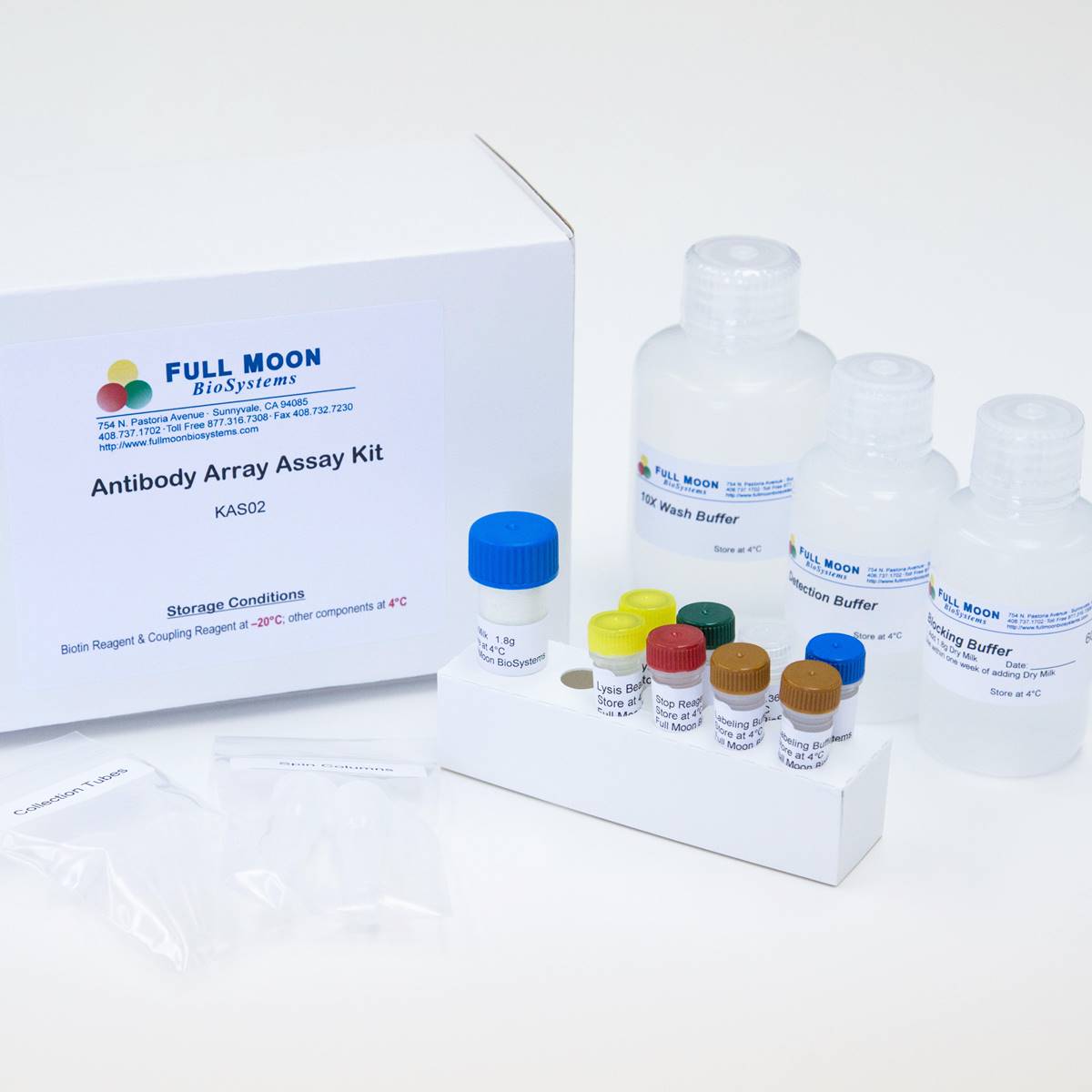
Complete Antibody Array Assay Service allows investigators to send research samples to our laboratory for analysis. There is no need to purchase the arrays and reagents and running the assays yourself. Simply select the array of your choice, and then send off the samples to our lab. This convenient hands-off approach offers quick turnaround and reliable results, saving you valuable time and resources. All assays will be performed by our highly trained scientists at our headquarter in Sunnyvale, California. Results are delivered by email in 1-3 weeks.
Cost: $2,380 per sample