TGF-beta Phospho Antibody Array
Transforming growth factor beta (TGF-β) signaling plays an important role in cell proliferation, differentiation, and repair of many organisms. The signaling process begins with a dimeric TGF-β superfamily ligand binding to a Type II receptor, which enlists a Type I receptor (TGFBR1) and phosphorylates its serine residues. The activated TGFBR1 then phosphorylates and activates cytoplasmic molecules Smad2 and Smad3 for the TGF-β pathway or Smad1/5/9 for the bone morphogenetic protein (BMP) pathway. The phosphorylation of Smads leads to formation of a hetero-oligomeric complex with Smad4, which then moves into the nucleus and interacts with transcription promoters/cofactors and regulates gene expression. TGF-β also participates in the regulation of non-Smad pathways including ERK1/2, JNK/SAPK, and p38 MAP kinase pathways.
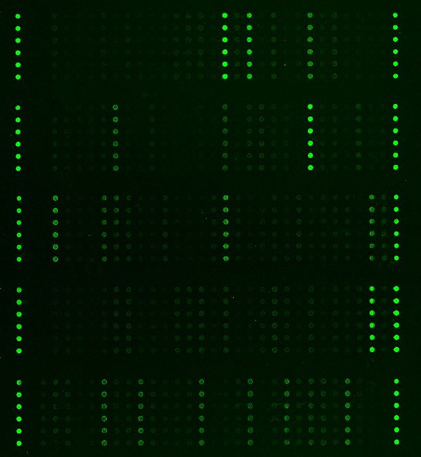
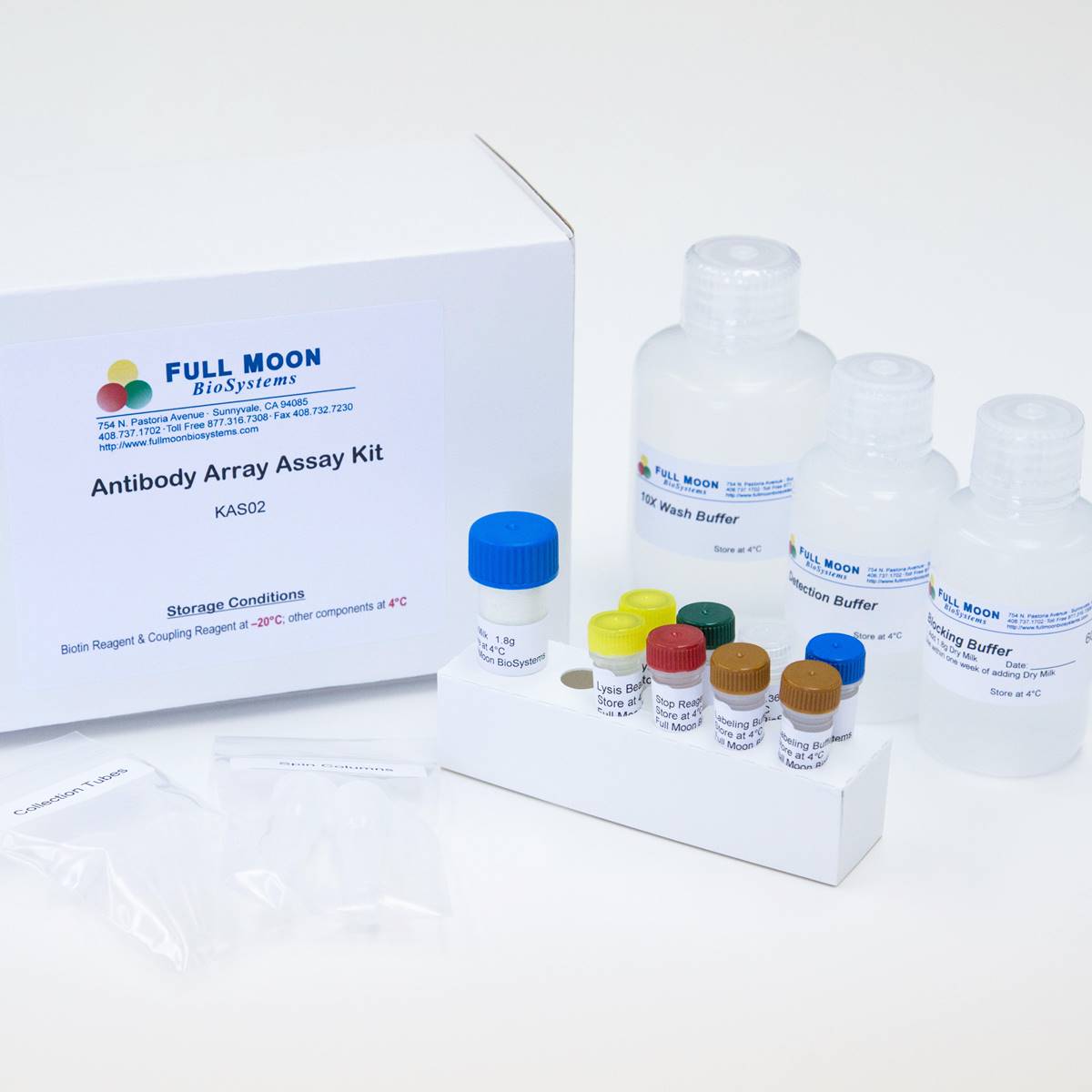
TGF-β (TGF-beta) Phospho Antibody Array is a high-throughput ELISA based antibody array for qualitative protein phosphorylation profiling. It is suitable for comparing normal samples to treated or diseased samples, and identifying candidate biomarkers. This array features site-specific and phospho-specific antibodies, allowing researchers to investigate serine/threonine and tyrosine phosphorylation at specific sites.
Key Features
- Site-specific phosphorylation profiling and screening with 176 antibodies
- Antibodies covalently immobilized on 3D polymer coated glass slide
- Fluorescent detection
Specifications
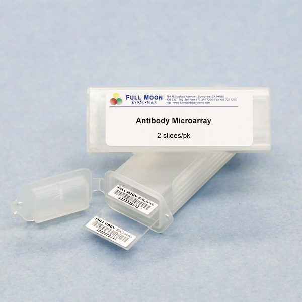
| Product Size: | 2 array slides per package for analyzing two samples (untreated vs. treated) |
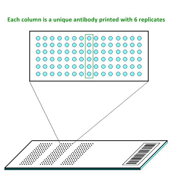
| Featured Antibodies: | 176 site-specific and phospho-specific antibodies; 6 replicates per antibody |
| Reactivity: | Human: 100% | Mouse: 97% | Rat: 87% |
| Suitable Sample Type: | Cell lysate | Tissue lysate |
| Detection Method: | Fluorescence | Compatible Scanners |
| Internal Controls: | beta-actin | GAPDH | Negative controls |
| Slide Dimensions: | 76 x 25 x 1 mm |
| Storage Condition: | 4°C for 6 months |
Product Details
AKT1 (Ser124), AKT1 (Ser246), AKT1 (Thr450), AKT1 (Thr72), AKT1 (Tyr474), AKT2 (Ser474), c-Abl (Tyr245), c-Abl (Tyr412), cofilin (Ser3), ERK1 (Thr202), ERK1 (Tyr204), ERK1/2, ERK3 (Ser189), ERK8 (Thr175/Tyr177), Gab2 (Tyr643), JNK1/2/3 (Thr183/Tyr185), JNKK, LIMK1 (Thr508), MAP3K1 (Thr1381), MKK3 (Ser189), MKK3 (Thr222), MKK6 (Ser207), mTOR (Ser2448), mTOR (Ser2481), mTOR (Thr2446), Myc (Ser373), Myc (Ser62), Myc (Thr358), Myc (Thr58), p38 MAPK (Thr180), p38 MAPK (Tyr182), p38 MAPK (Tyr322), PAK1 (Ser204), PAK1 (Thr212), PAK1/2 (Ser199), PAK1/2/3 (Ser141), PAK1/2/3 (Thr423/402/421), PAK2 (Ser192), PAK2 (Ser20), PAK3 (Ser154), PI3K-p85-a (Tyr607), PI3K-p85-a/g (Tyr467/199), PKC alpha (Tyr657), PKC alpha/beta II (Thr638), PKC beta (Ser661), PKC delta (Ser645), PKC delta (Thr505), PKC epsilon (Ser729), PKC theta (Ser676), PKC theta (Thr538), PKC zeta (Thr410), PKC zeta (Thr560), PP2A-alpha (Tyr307), Rac1/cdc42 (Ser71), Ras-GRF1 (Ser916), Rho/Rac GEF2 (Ser885), S6 Ribosomal Protein (Ser235), S6K, SAPK (Thr183), SAPK (Tyr185), SEK1 (Ser80), SEK1 (Thr261), Shc (Tyr349), Shc (Tyr427), Smad1 (Ser187), Smad1 (Ser465), Smad1/5/9, Smad2 (Ser250), Smad2 (Ser467), Smad2 (Thr220), Smad2/3 (Thr8), Smad3 (Ser204), Smad3 (Ser208), Smad3 (Ser213), Smad3 (Ser425), Smad3 (Thr179), Smad4, SP1 (Thr739), TAK1 (Thr184), TGF alpha, TGFB1, TGFB2, TGFB3, TGFBR2
The ELISA based TGF-beta Signaling Phospho Antibody Array platform involves four major steps:
- Protein extraction with non-denaturing lysis buffer
- Biotinylation of protein samples
- Incubation of labeled samples with antibody array
- Detection by dye conjugated streptavidin

Ciullo A, Li L, Non-coding RNA yREX3 from human extracellular vesicles exerts macrophage-mediated cardioprotection via a novel gene-methylating mechanism, Eur Heart J. 2024 Jun 12:ehae357. doi: 10.1093/eurheartj/ehae357
Kattih B, Boeckling F, Single-nuclear transcriptome profiling identifies persistent fibroblast activation in hypertrophic and failing human hearts of patients with longstanding disease, Cardiovasc Res. 2023 Aug 30;cvad140. doi: 10.1093/cvr/cvad140
Müller A., Lozoya M., Farnesol Inhibits PI3 Kinase Signaling and Inflammatory Gene Expression in Primary Human Renal Epithelial Cells. Biomedicines 2023. 11:3322. doi.org/10.3390/biomedicines11123322
Phillips PL, Reyes L. Porphyromonas gingivalis Type 9 Secretion System Promotes Dysregulation of Vascular Smooth Muscle Cell Plasticity with Perturbed TGF-β/Smad Signaling. Microbiologyopen. 2025 Oct;14(5):e70044. doi: 10.1002/mbo3.70044
Santerre K, Ghio SC, TGF-b-Mediated Modulation of Cell-Cell Interactions in Postconfluent Maturing Corneal Endothelial Cells, Invest Ophthalmol Vis Sci. 2022 Oct 3;63(11):3. doi: 10.1167/iovs.63.11.3
Services
If you don’t have access to a microarray, send the finished arrays to our lab for scanning. Raw scan images are delivered in tiff format.
Cost: Free
Array Image Quantification and Analysis Service includes data extraction, data organization and analysis of the array images obtained through our array scanning service.
Cost: $210 per slide
Complete Antibody Array Assay Service allows investigators to send research samples to our laboratory for analysis. There is no need to purchase the arrays and reagents and running the assays yourself. Simply select the array of your choice, and then send off the samples to our lab. This convenient hands-off approach offers quick turnaround and reliable results, saving you valuable time and resources. All assays will be performed by our highly trained scientists at our headquarter in Sunnyvale, California. Results are delivered by email in 1-3 weeks.
Cost: $1,515 per sample