Insulin Receptor Phospho Antibody Array
Insulin Receptor Phospho Antibody Array is a high-throughput ELISA based antibody array for qualitative protein phosphorylation profiling. It is suitable for comparing normal samples to treated or diseased samples, and identifying candidate biomarkers. This array features site-specific and phospho-specific antibodies, allowing researchers to study tyrosine phosphorylation and serine/threonine phosphorylation at specific sites.
Key Features
- Site-specific phosphorylation profiling and screening
- Suitable samples include: cell lysates; frozen or FFPE tissue lysates
- Glass based array with high specificity and low background
- Antibodies covalently attached to 3D polymer coated glass surface
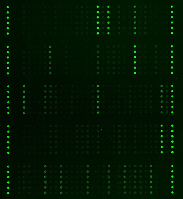
- Sensitive fluorescent detection
Specifications
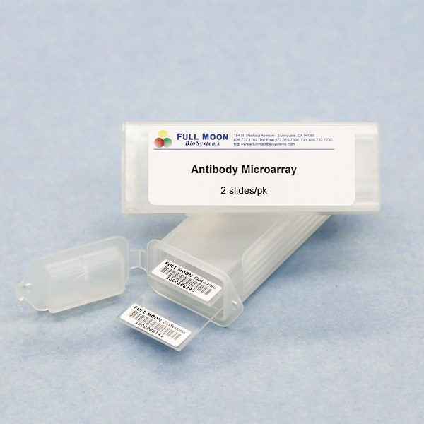
| Product Size: | 2 array slides per package for analyzing two samples (untreated vs. treated) |
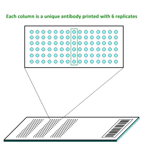
| Featured Antibodies: | 219 site-specific and phospho-specific antibodies; 6 replicates per antibody |
| Reactivity: | Human: 100% | Mouse: 97% | Rat: 84% |
| Suitable Sample Type: | Cell lysate | Tissue lysate |
| Detection Method: | Fluorescence | Compatible Scanners |
| Internal Controls: | beta-actin | GAPDH | Negative controls |
| Slide Dimensions: | 76 x 25 x 1 mm |
| Storage Condition: | 4°C for 6 months |
PRODUCT DETAILS
4E-BP1 (Ser65), 4E-BP1 (Thr36), 4E-BP1 (Thr45), 4E-BP1 (Thr70), AKT (Ser473), AKT (Thr308), AKT (Tyr326), AKT1 (Ser124), AKT1 (Ser246), AKT1 (Thr450), AKT1 (Thr72), AKT1 (Tyr474), AKT1S1 (Thr246), AKT2 (Ser474), AMPK1 (Thr174), AMPK1/AMPK2 (Ser485/491), AMPKbeta1 (Ser182), ATP-Citrate Lyase (Ser454), BAD (Ser112), BAD (Ser134), BAD (Ser136), BAD (Ser155), BAD (Ser91/128), Caveolin-1 (Tyr14), CBL (Tyr700), CBL (Tyr774), c-Raf (Ser296), c-Raf (Ser43), CrkII (Tyr221), CrkL (Tyr207), eIF2a (Ser51), eIF4B (Ser422), eIF4E (Ser209), eIF4G (Ser1108), ERK3 (Ser189), ERK8 (Thr175/Tyr177), FKHR (Ser256), FKHR (Ser319), FKHRL1 (Ser253), FOXO1/3/4-PAN (Thr24/32), FOXO1A (Ser329), FOXO1A/3A (Ser322/325), Gab1 (Tyr627), Gab1 (Tyr659), GRB2 (Ser159), GRB10 (Tyr67), GSK3a (Ser21), GSK3a/b (Tyr216/279), GSK3b (Ser9), HSL (Ser552/563), HSL (Ser554), IKKa (Thr23), IKKa/b (Ser180/181), IKKb (Tyr188), IKKb (Tyr199), IKKg (Ser31), IKKg (Ser85), IR (Tyr1355), IR (Tyr1361), IRS-1 (Ser1101), IRS-1 (Ser307), IRS-1 (Ser312), IRS-1 (Ser323), IRS-1 (Ser612), IRS-1 (Ser636), IRS-1 (Ser639), IRS-1 (Ser794), LKB1 (Ser428), LKB1 (Thr189), MEK1 (Ser217), MEK1 (Ser221), MEK1 (Ser298), MEK1 (Thr286), MEK1 (Thr291), MEK2 (Thr394), mTOR (Ser2448), mTOR (Ser2481), mTOR (Thr2446), p44/42MAPK (Thr202), ERK1-p44/42MAPK (Tyr204), p70S6K beta (Ser423), p70S6K (Ser371), p70S6K (Ser411), p70S6K (Ser418), p70S6K (Ser424), p70S6K (Thr229), p70S6K (Thr389), p70S6K (Thr421), PDK1 (Ser241), PI3K-p85-a (Tyr607), PI3K-p85-a/g (Tyr467/Tyr199), PKA CAT (Thr197), PKC pan activation site, PKC theta (Ser676), PKC theta (Thr538), PKC zeta (Thr410), PKC zeta (Thr560), PP1a (Thr320), PP2A-a (Tyr307), PTEN (Ser370), PTEN (Ser380), PTEN (Ser380/Thr382/Thr383), PTPRA (Tyr798), RASE, RASF4, Ras-GRF1 (Ser916), Shc (Tyr349), Shc (Tyr427), SHP-2 (Tyr542), SHP-2 (Tyr580), TNFR1, TNFR2, Tuberin (Ser939), Tuberin (Thr1462)
The ELISA based Insuline Receptor Phospho Antibody Array platform involves four major steps:
- Protein extraction with non-denaturing lysis buffer
- Biotinylation of protein samples
- Incubation of labeled samples with antibody array
- Detection by dye conjugated streptavidin

Fischer HJ, Sie C, The Insulin Receptor Plays a Critical Role in T Cell Function and Adaptive Immunity, J Immunol. 2017 Mar 1;198(5):1910-1920
He H, Zong Y, Sensing the Insulin Signaling Pathway with an Antibody Array, Proteomics Clinical Applications, 2009;3:1440–1450
Kwan SH, de Mejía EG. Peptides from adzuki bean and soybean improved insulin-AKT signaling-related pathways in healthy and insulin-resistant states in human liver cells. Mol Nutr Food Res. 2025. doi:10.1002/mnfr.70285
Luna-Vital D, Weiss M, Anthocyanins from Purple Corn Ameliorated TNF-α-Induced Inflammation and Insulin Resistance in 3T3-L1 Adipocytes via Activation of Insulin Signaling and Enhanced GLUT4 Translocation, Mol Nutr Food Res. 2017 July 31, doi: 10.1002/mnfr.201700362
Rebollo‐Hernanz M, Zhang Q, Cocoa Shell Aqueous Phenolic Extract Preserves Mitochondrial Function and Insulin Sensitivity by Attenuating Inflammation Between Macrophages and Adipocytes in vitro, Mol Nutr Food Res. 2019 Apr 24:e1801413
Rozenberg K, Smirin P, Insulin-sensitizing and insulin-mimetic activities of Sarcopoterium spinosum extract, J Ethnopharmacology, August 8 2014, 155(1): 362-372
Sweidan R, de Souza A, Metformin attenuates cardiac deficiencies and repairs hepatic insulin signaling in a rat model of acute food insecurity followed by recovery, American Physiology Summit 2023 Meeting Abstracts, May 2023, https://doi.org/10.1152/physiol.2023.38.S1.5733203
Services
If you don’t have access to a microarray, send the finished arrays to our lab for scanning. Raw scan images are delivered in tiff format.
Cost: Free
Array Image Quantification and Analysis Service includes data extraction, data organization and analysis of the array images obtained through our array scanning service.
Cost: $260 per slide
Complete Antibody Array Assay Service allows investigators to send research samples to our laboratory for analysis. There is no need to purchase the arrays and reagents and running the assays yourself. Simply select the array of your choice, and then send off the samples to our lab. This convenient hands-off approach offers quick turnaround and reliable results, saving you valuable time and resources. All assays will be performed by our highly trained scientists at our headquarter in Sunnyvale, California. Results are delivered by email in 1-3 weeks.
Cost: $1,565 per sample