Cell Signaling Phospho Antibody Array
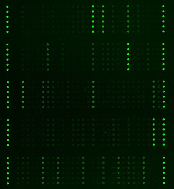
The Cell Signaling Phospho Antibody Array is a high-throughput ELISA based antibody array for qualitative profiling and screening of candidate biomarkers from 16 cell signaling pathways, including PI3K/AKT signaling, apoptosis, autophagy, cell cycle, ErbB, focal adhension, MAPK, p53 signaling, VEGF and more. It is designed to identify total protein changes and phosphorylation changes by comparing normal samples to treated/diseased samples. This array features site-specific and phospho-specific antibodies for studies of phosphorylation events at specific phospho sites.
Key Features
- 304 antibodies from 16 cell signaling pathways
- Site-specific phosphorylation profiling and screening
- Antibodies covalently immobilized on 3D polymer coated glass slide
- Fluorescent detection
Specifications
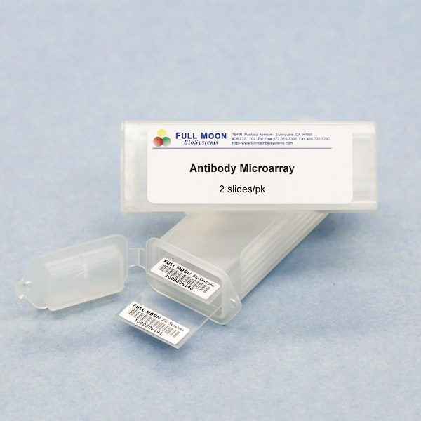
| Product Size: | 2 array slides per package for analyzing two samples (untreated vs. treated) |
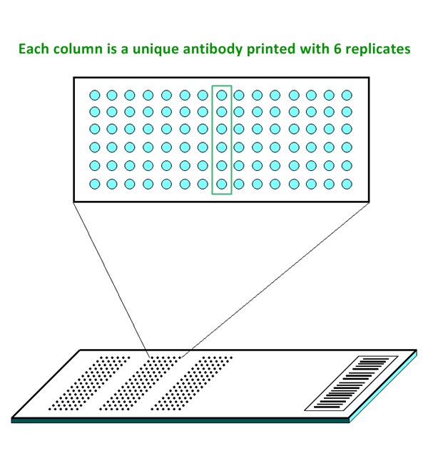
| Featured Antibodies: | 304 site-specific and phospho-specific antibodies; 6 replicates per antibody |
| Reactivity: | Human: 99% | Mouse: 87% | Rat: 66% |
| Suitable Sample Type: | Cell lysate | Tissue lysate |
| Detection Method: | Fluorescence | Compatible Scanners |
| Internal Controls: | beta-actin | GAPDH | Negative controls |
| Slide Dimensions: | 76 x 25 x 1 mm |
| Storage Condition: | 4°C for 6 months |
Product Details
PI3K/AKT
AKT1, AKT2, BAD, BCL2, CASP9, CCND1, CCNE1, CDK2, CKDN1A, CHUK, CREB1, EGFR1, EIF4E, EIF4EBP1, ERBB2, FOXO3, GSK3B, HSP90AB1, IGF1R, IKBKB, IKBKG, IL4R, IRS1, ITGB3, JAK1, JAK2, KDR, MAP2K1, MDM2, MET, MTOR, MYC, NFKB1, NOS3, NTRK2, PDGFRB, PDK1, PPP2CA, PRKAA1, PTEN, PTK2, RAF1, RELA, RPS6KB1, STK11, SYK, TP53, TSC2, YWHAQ
AMPK
ACACA, AKT1, AKT1S1, AKT2, CCND1, CREB1, EEF2, EEF2K, EIF4EBP1, FOXO1, FOXO3, IFG1R, IRS1, MAP3K7, MTOR, PDPK1, PPARG, PPP2CA, PRKAA1, RPS6KB1, SREBF1, STK11, TSC2
Apoptosis
ACTB, ACTG1, AKT1, AKT2, BAD, BAX, BCL2, BID, CASP3, CASP8, CASP9, CHUK, EIF2S1, FADD, FOS, IKBKB, IKBKG, LMNA, MAP2K1, MAP3K5, MAPK9, NFKB1, NFKB1A, PDPK1, RAF1, RELA, TP53
Autophagy
AKT1, AKT1S1, AKT2, BAD, BCL2, BECN1, EIF2S1, IGF1R, IRS1, MAP2K1, MAP3K7, MAPK9, MTOR, PDPK1, PPP2CA, PRKAA1, PTEN, RAF1, RPS6KB1, STK11, TSC2
Cell Cycle
ABL1, CCND1, CCNE1, CDC25C, CDK2, CDKNIA, CHEK1, CHEK2, GSK3B, HDAC1, HDAC2, MDM2, MYC, PLK1, RB1, SMAD2, SMAD3, TP53, YWHAQ
ErbB
ABL1, AKT1, AKT2, BAD, BRAF, CAMK2A, CAMK2B, CAMK2D, CAMK2G, CBL, CDKN1A, CRK, EGFR, EIF4EBP1, ELK1, ERBB2, GAB1, GSK3B, MAP2K1, MAP2K4, MAPK9, MTOR, MYC, PAK1, PLCG1, PTK2, RAF1, RPS6KB1, SHC1, SRC, STAT5A, STAT5B ,
Focal Adhesion
ACTB, ACTG1, AKT1, AKT2, BAD, BCL2, BRAF, CCND1, CRK, CTNNB1, EGFR, ELK1, ERBB2, GSK3B, IGF1R, ITGB3, KDR, MAP2K1, MAKP9, MET, PAK1, PDGFRB, PDPK1, PPP1CA, PTEN, PTK2, RAF1, RASGRF1, SHC1, SRC, VASP
Insulin
ACACA, AKT1, AKT2, BAD, BRAF, CALM1, CBL, CRK, EIF4E, EIF4EBP1, ELK1, FOXO1, GSK3B, IKBKB, IRS1, MAK2K1, MAPK9, MTOR, PDPK1, PPP1CA, PRKAA1, RAF1, RPS6KB1, SHC1, SREBF1, TSC2 ,
JAK-STAT
AKT1, AKT2, BCL2, CCND1, CDKN1A, EGFR, IFNGR1, IL10RA, IL4R, JAK1, JAK2, MTOR, MYC, PDGFRB, PTPN11, RAF1, STAT1, STAT2, STAT3, STAT4, STAT5A, STAT5B, STAT6
MAPK
AKT1, AKT2, BRAF, CHUK, CRK, EGFR, ELK1, ERBB2, FOS, HSPB1, IGR1R, IKBKG, KDR, MAP2K1, MAP2K4, MAP3K5, MAP3K7, MAPK14, MAPK9, MAPT, MET, MYC, NFKB1, NTRK2, PAK1, PDFGRB, RAF1, RASGRF1, RELA, RELB, RPS6KA1, RPS6KA2, RPS6KA3, RPS6KA5, SRF, TP53, CASP3
mTOR
AKT1, AKT1S1, AKT2, BRAF, CHUK, EIF4E, EIF4EBP1, GSK3B, IFG1R, IKBKB, IRS1, MAP2K1, MTOR, PDPK1, PRKAA1, PTEN, RAF1, RPS6KA1, RPS6KA2, RPS6KA3, RPS6KB1, STK11, TSC2, WNT1
NF-kappa B
BCL2, BLNK, CHUK, IKBKB, IKBKG, LCK, LYN, MAP3K7, NFKB1, NFKB1A, PLCG1, RELA, RELB, SYK, ZAP70
p53
BAX, BCL2, BID, CASP8, CASP9, CCND1, CCNE1, CDK2, CDKNIA, CHEK1, CHEK2, MDM2, PTEN, TP53, TP73, TSC2, CASP3, CASP8
Ras
ABL1, AKT1, AKT2, BAD, CALM1, CHUK, EGFR, ELK1, GAB1, GAB2, GRIN1, IGF1R, IKBKB, IKBKG, KDR, MAP2K1, MAPK9, MET, NFKB1, PAK1, PDFGRB, PLCG1, PTPN11, RAF1, RASGRF1, RELA, SHC1, ZAP70
TGF-beta
MYC, PPP2CA, RPS6KB1, SMAD1, SMAD2, SMAD3, TGFBR1, TGFBR2
VEGF
AKT1, AKT2, BAD, CASP9, HSPB1, MAP2K1, MAPK14, NOS3, PLCG1, PTK2, RAF1, SRC, VEGFR2
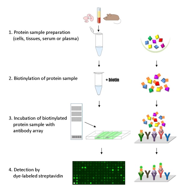
The ELISA based Cell Signaling Phospho Antibody Array platform involves four major steps:
- Protein extraction with non-denaturing lysis buffer
- Biotinylation of protein samples
- Incubation of labeled samples with antibody array
- Detection by dye conjugated streptavidin
Ashraf, S, Taegtmeyer H, Prolonged cardiac NR4A2 activation causes dilated cardiomyopathy in mice, Basic Res Cardiol. 2022 Jul 1;117(1):33. doi: 10.1007/s00395-022-00942-7
Chalise U, Becirovic-Agic, MMP-12 polarizes the neutrophil signalome towards an apoptotic signature, J. Proteomics. 2022. https://doi.org/10.1016/j.jprot.2022.104636
Choe YJ, Min JY, Heterotypic cell-in-cell structures between cancer and NK cells is associated with enhanced anti-cancer drug resistance, iScience. 2002 Aug 27, 105017. https://doi.org/10.1016/j.isci.2022.105017
Choi J, Hwang J, Inhibition of miR-4284 could reduce apoptosis and neuroinflammation by targeting APBA1/JAK1/STAT3 signaling in Alzheimer’s disease. Cell Biosci 15, 151 (2025). https://doi.org/10.1186/s13578-025-01498-4
Jungholm O, Trkulja C, Novel druggable space in human KRAS G13D discovered using structural bioinformatics and a P-loop targeting monoclonal antibody. Sci Rep. 2024, 3;14(1):19656. doi: 10.1038/s41598-024-70217-9.
Sun X, Chen M, Knockdown of KIF15 promotes cell apoptosis by activating crosstalk of multiple pathways in ovarian cancer: bioinformatic and experimental analysis, Int J Clin Exp Pathol. 2021 Feb 1;14(2):267-291
Wang H, Ge L, 3,4,5-Tri-O-caffeoylquinic acid methyl ester isolated from Lonicera japonica Thunb. Flower buds facilitates hepatitis B virus replication in HepG2.2.15 cells, Food Chem Toxcol. 2020 March 7. doi: 10.1016/j.fct.2020.111250
Ye Y, Wang H, Polygalasaponin F treats mice with pneumonia induced by influenza virus, Inflammopharmacology. 2019 Aug 24. doi: 10.1007/s10787-019-00633-1
Services
If you don’t have access to a microarray, send the finished arrays to our lab for scanning. Raw scan images are delivered in tiff format.
Cost: Free
Array Image Quantification and Analysis Service includes data extraction, data organization and analysis of the array images obtained through our array scanning service.
Cost: $310 per slide
Complete Antibody Array Assay Service allows investigators to send research samples to our laboratory for analysis. There is no need to purchase the arrays and reagents and running the assays yourself. Simply select the array of your choice, and then send off the samples to our lab. This convenient hands-off approach offers quick turnaround and reliable results, saving you valuable time and resources. All assays will be performed by our highly trained scientists at our headquarter in Sunnyvale, California. Results are delivered by email in 1-3 weeks.
Cost: $1,780 per sample