mTOR Phospho Antibody Array
The mechanistic target of rapamycin (mTOR) is a serine-threonine protein kinase that regulates cell growth, cell proliferation, protein synthesis, and autophagy. Abnormal mTOR signaling has been associated with neurological disorders, cancer, and diabetes. mTOR forms two distinct complexes: mTOR Complex 1 (mTORC1) and mTOR Complex 2 (mTORC2). mTOR binds with the regulatory protein raptor and other binding proteins to form mTORC1 while it binds with regulatory protein rictor and other proteins to form mTORC2. mTORC1 phosphorylates p70S6K and eIF4E to promote mRNA translation and protein synthesis. Growth factors such as insulin trigger the AKT pathway, which signals downstream to mTORC1 to promote cell growth and inhibit autophagy. Oppositely, nutrient deprivation and low energy levels activate the AMPK pathway, which inhibits mTORC1, thus restricting cell growth and promoting autophagy. mTORC2 can affect metabolism and cell survival through phosphorylation of AKT. mTORC2 regulates the cytoskeleton through PKC alpha activation and also influences ion transport.
mTOR Phospho Antibody Array is a high-throughput ELISA based antibody array for qualitative protein phosphorylation profiling. It is suitable for comparing normal samples to treated or diseased samples, and identifying candidate biomarkers. This array features site-specific and phospho-specific antibodies, allowing researchers to study tyrosine, serine, and threonine phosphorylation at specific sites.
Key Features
- Site-specific phosphorylation profiling and screening with 138 antibodies
- Antibodies covalently immobilized on 3D polymer coated glass slide
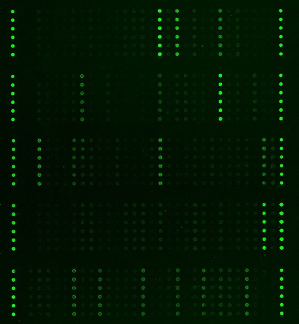
- Fluorescent detection
Specifications
| Product Size: | 2 array slides per package for two samples (untreated vs. treated) |
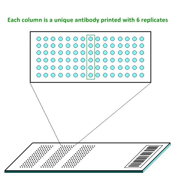
| Featured Antibodies: | 138 site-specific and phospho-specific antibodies; 6 replicates per antibody |
| Reactivity: | Human: 100% | Mouse: 97% | Rat: 83% |
| Suitable Sample Type: | Cell lysate | Tissue lysate |
| Detection Method: | Fluorescence | Compatible Scanners |
| Internal Controls: | Positive controls: beta-actin | GAPDH | Negative controls |
| Slide dimensions: | 76 x 25 x 1 mm |
| Storage Condition: | 4°C for 6 months |
Product Details
4E-BP1 (Ser65), 4E-BP1 (Thr36), 4E-BP1 (Thr45), 4E-BP1 (Thr70), AKT1 (Ser124), AKT1 (Ser246), AKT1 (Thr450), AKT1 (Thr72), AKT1 (Tyr474), AKT1S1 (Thr246), AKT2 (Ser474), AKT (Ser473), AKT (Thr308), AKT (Tyr326), AMPK1/AMPK2 (Ser485/491), AMPK1 (Thr174), AMPKbeta1 (Ser182), BAD (Ser112), BAD (Ser134), BAD (Ser136), BAD (Ser155), BAD (Ser91/128), eIF2A (Ser51), eIF4B (Ser422), eIF4E (Ser209), eIF4G (Ser1108), ERK1/2, ERK1 (Thr202), ERK1 (Tyr204), ERK3 (Ser18), ERK8 (Thr175/Tyr177), GSK3a (Ser21), GSK3a-b (Tyr216/270), GSK3b (Ser9), IR (Tyr1355), IR (Tyr1361), Mnk1 (Thr385), mTOR (Ser2448), mTOR (Ser2481), mTOR (Thr2446), P70S6K beta (Ser423), P70S6K (Ser371), P70S6K (Ser411), P70S6K (Ser418), P70S6K (Ser424), P70S6K (Thr229), P70S6K (Thr389), P70S6K (Thr421), P90RSK (Ser380), P90RSK (Thr359/Ser363), P90RSK (Thr573), PDK1 (Ser241), PFKFB2 (Ser483), PI3K-p85-a (Tyr607), PI3K-p85-a/g (Tyr467/Tyr199), PIP5K (Ser307), PKC alpha (Tyr657), PKC alpha/beta II (Thr638), PP2A-alpha (Tyr307), PPAR-b (Thr1457), PPAR-g (Ser112), PTEN (Ser370), PTEN (Ser380), PTEN (Ser380/Thr382/Thr383), Rho/Rac GEF2 (Ser885), RSK1/2/3/4 (Ser221/227/218/232), Tuberin (Ser939), Tuberin (Thr1462)
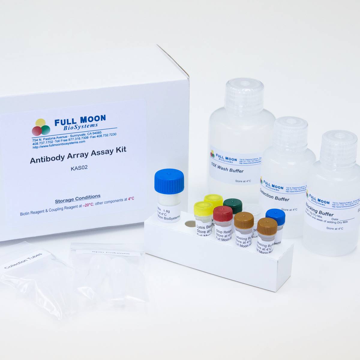
The ELISA based mTOR Pathway Phospho Antibody Array platform involves four major steps:
- Protein extraction with non-denaturing lysis buffer
- Biotinylation of protein samples
- Incubation of labeled samples with antibody array
- Detection by dye conjugated streptavidin

![]() GAL File (To download, right click on the file name, then choose “Save target as”)
GAL File (To download, right click on the file name, then choose “Save target as”)
Alqurashi N, Hashimi SM, Dual mTOR/PI3K inhibitor NVP‑BEZ235 arrests colorectal cancer cell growth and displays differential inhibition of 4E‑BP1, Oncol Rep, 2018 May 22, https://doi.org/10.3892/or.2018.6457
Arsenault, R, Napper, S, Salmonella enterica Typhimurium infection causes metabolic changes in chicken muscle involving AMPK, fatty acid and insulin/mTOR signaling, Vet Res. 2013, 44:35
Majocha MR, Jackson DE, Resf1 is a compound G4 quadruplex-associated tumor suppressor for triple negative breast cancer. PLoS Genet. 2024 May 9;20(5):e1011236. doi: 10.1371/journal.pgen.1011236.
Wang H, Brown J, et al.: Convergence of the Mammalian Target of Rapamycin Complex 1- and Glycogen Synthase Kinase 3-b–Signaling Pathways Regulates the Innate Inflammatory Response, J. Immunol. 2011, 186:5217-5226
Services
If you don’t have access to a microarray, send the finished arrays to our lab for scanning. Raw scan images are delivered in tiff format.
Cost: Free
Array Image Quantification and Analysis Service includes data extraction, data organization and analysis of the array images obtained through our array scanning service.
Cost: $210 per slide
Complete Antibody Array Assay Service allows investigators to send research samples to our laboratory for analysis. There is no need to purchase the arrays and reagents and running the assays yourself. Simply select the array of your choice, and then send off the samples to our lab. This convenient hands-off approach offers quick turnaround and reliable results, saving you valuable time and resources. All assays will be performed by our highly trained scientists at our headquarter in Sunnyvale, California. Results are delivered by email in 1-3 weeks.
Cost: $1,410 per sample