
Jak/Stat Phospho Antibody Array
Jak/Stat Phospho Antibody Array is a high-throughput ELISA based antibody array for qualitative protein phosphorylation profiling. It is suitable for comparing normal samples to treated or diseased samples and identifying candidate biomarkers. This array features site-specific and phospho-specific antibodies, allowing researchers to study tyrosine, serine, and threonine phosphorylation at specific sites.
Key Features
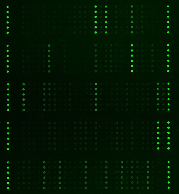
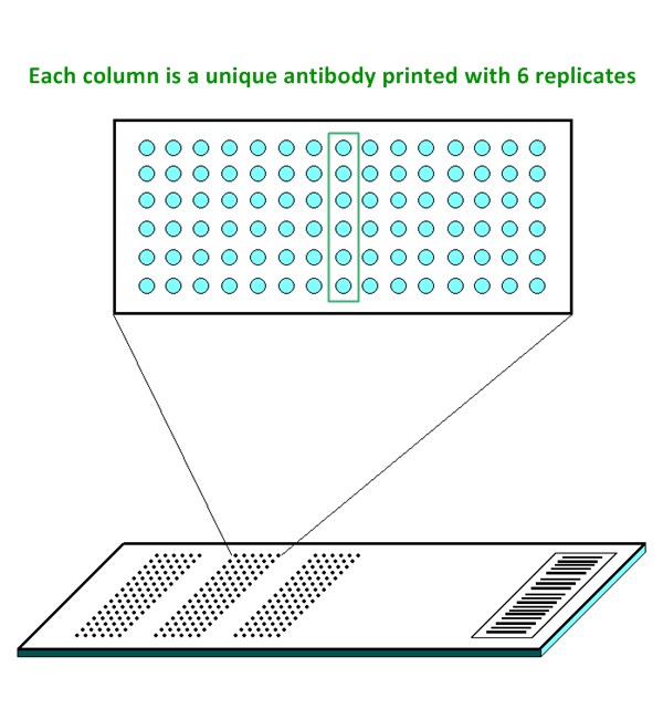
- Site-specific phosphorylation profiling and screening with 42 antibodies
- Antibodies covalently immobilized on 3D polymer coated glass slide
- Fluorescent detection
Specifications
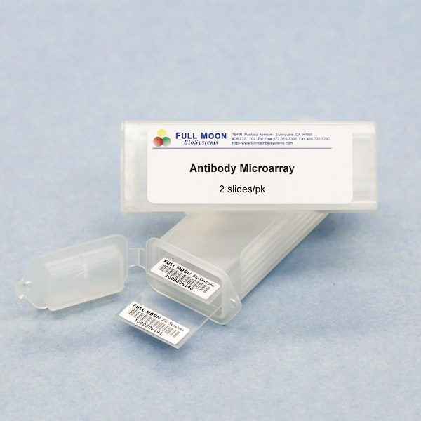
| Product Size: | 2 array slides per package for analyzing two samples (untreated vs. treated) |
| Featured Antibodies: | 42 site-specific and phospho-specific antibodies; 6 replicates per antibody |
| Reactivity: | Human: 100% | Mouse: 93% | Rat: 86% |
| Suitable Sample Type: | Cell lysate | Tissue lysate |
| Detection Method: | Fluorescence | Compatible Scanners |
| Internal Controls: | beta-actin | GAPDH | Negative controls |
| Slide Dimensions: | 76 x 25 x 1 mm |
| Storage Condition: | 4°C for 6 months |
Product Details
JAK1 (Tyr1022), JAK2 (Tyr1007), JAK2 (Tyr221), MEK1 (Ser217), MEK1 (Ser221), MEK1 (Thr291), MEK2 (Thr394), p44/42 MAPK (Thr202), p44/42 MAPK (Tyr204), Raf1 (Ser259), Raf1 (Ser338), STAT1 (Ser727), STAT1 (Tyr701), STAT3 (Ser727), STAT3 (Tyr705), STAT4 (Tyr693), STAT5A (Ser780), STAT5A (Tyr694), STAT6 (Thr645), STAT6 (Tyr641), TYK2 (Tyr1054)
The ELISA based Jak/Stat Phospho Antibody Array platform involves four major steps:
- Protein extraction with non-denaturing lysis buffer
- Biotinylation of protein samples
- Incubation of labeled samples with antibody array
- Detection by dye conjugated streptavidin

Fierros-Zárate G, Olvera C, Bovine Interferon-Tau Activates Type I interferon-Associated Janus-signal Transducer in HPV16-positive Tumor Cell, J Cancer 2020; 11(16):4754-4761. doi:10.7150/jca.33527
Heneghan AF, Pierre JF, IL-25 Improves IgA Levels During Parenteral Nutrition Through the JAK-STAT Pathway, Annals of Surgery 2013 Dec;258(6):1065-71
Pierre JF, Heneghan AF, Cranberry Proanthocyanidins Improve Intestinal sIgA During Elemental Enteral Nutrition, Journal of Parenteral & Enteral Nutrition 2013, doi: 10.1177/0148607112473654
Rayasam a, Kijak J, CXCL13 Expressed on Inflamed Cerebral Blood Vessels Recruit IL-21 Producing TFH Cells to Damage Neurons Following Stroke, J Neuroinflammation 2021, Preprint
Additional Services
If you don’t have access to a microarray, send the finished arrays to our lab for scanning. Raw scan images are delivered in tiff format.
Cost: Free
Array Image Quantification and Analysis Service includes data extraction, data organization and analysis of the array images obtained through our array scanning service.
Cost: $160 per slide
Complete Antibody Array Assay Service allows investigators to send research samples to our laboratory for analysis. There is no need to purchase the arrays and reagents and running the assays yourself. Simply select the array of your choice, and then send off the samples to our lab. This convenient hands-off approach offers quick turnaround and reliable results, saving you valuable time and resources. All assays will be performed by our highly trained scientists at our headquarter in Sunnyvale, California. Results are delivered by email in 1-3 weeks.
Cost: $1,275 per sample