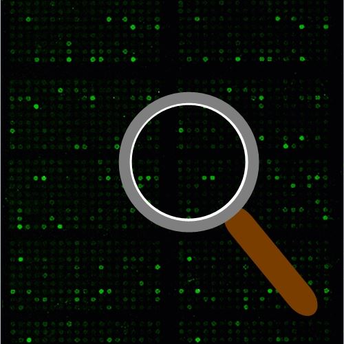
Cytoskeleton Phospho Antibody Array
Cytoskeleton Phospho Antibody Array is a high-throughput ELISA based antibody array for qualitative protein phosphorylation profiling. It is designed for studying key proteins for cytoskeletal signaling and regulation, suitable for comparing normal samples to treated or diseased samples and identifying candidate biomarkers. This array features site-specific and phospho-specific antibodies, allowing researchers to study tyrosine and serine/threonine phosphorylation at specific sites.
Key Features
- Site-specific phosphorylation profiling and screening with 141 antibodies
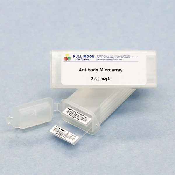
- Antibodies covalently immobilized on 3D polymer coated glass slide
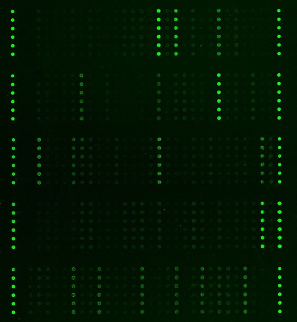
- Fluorescent detection
Specifications
| Product Size: | 2 array slides per package for analyzing two samples (untreated vs. treated) |
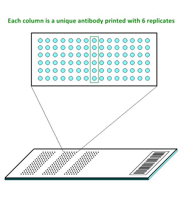
| Featured Antibodies: | 141 site-specific and phospho-specific antibodies; 6 replicates per antibody |
| Reactivity: | Human: 100% | Mouse: 100% | Rat: 81% |
| Suitable Sample Type: | Cell lysate | Tissue lysate |
| Detection Method: | Fluorescence | Compatible Scanners |
| Internal Controls: | beta-actin | GAPDH | Negative controls |
| Slide Dimensions: | 76 x 25 x 1 mm |
| Storage Condition: | 4°C for 6 months |
Product Details
Actin Pan (a/b/g) (Tyr55/53), Actin alpha-2/3, Actin-alpha1 sk muscle, ACTN1, Calmodulin (Thr79/Ser81), CaMK1-alpha (Thr177), CaMK1-beta, CaMK2 (Thr286), CaMK2 (Thr305), CaMK2-b/g/d (Thr287), CaMK4 (Thr196/200), CAMK5, Cofilin (Ser3), Cortactin (Tyr421), Cortactin (Tyr466), c-Raf (Ser296), c-Raf (Ser43), CrkII (Tyr221), CrkL (Tyr207), ERK1/2, ERK3 (Ser189), ERK8 (Thr175/Tyr177), Ezrin (Thr566), Ezrin (Tyr353), Ezrin (Tyr478), FAK (Ser910), FAK (Tyr397), FAK (Tyr407), FAK (Tyr576), FAK (Tyr861), FAK (Tyr925), Filamin A (Ser2152), GRB2 (Ser159), GTPase activating protein (Ser387), LIMK1 (Thr508), MEK1 (Ser217), MEK1 (Ser221), MEK1 (Thr291), MEK1 (Ser298), MEK1 (Thr286), MEK2 (Thr394), MEKKK1, MEKKK4, Merlin (Ser10), Merlin (Ser518), MKK3 (Ser189), MKK3/MAP2K3 (Thr222), MKK6 (Se207), MKK7/MAP2K7 (Ser271), MKK7/MAP2K7 (Thr275), Myosin regulatory light chain 2 (Ser18), NCK2, p130Cas (Tyr165), p130Cas (Tyr410), p44/42 MAPK (Thr202), p44/42 MAPK (Tyr204), Paxillin (Tyr118), Paxillin (Tyr31), PI3K-p85-alpha (Tyr607), PI3K-p85-a/g (Tyr467/Tyr199), PIP5K, PIP5K (Ser307), PKA CAT (Thr197), PKC alpha (Tyr657), PKC alpha/beta II (Thr638), PKC pan activation site, PLC beta (Ser1105), PLC beta3 (Ser537), Rac1/cdc42 (Ser71), Rho/Rac GEF (Ser885), Src (Ser75), Src (Tyr216), Src (Tyr418), Src (Tyr529), VASP (Ser157), VASP (Ser238), WASP (Tyr290), WAVE1 (Tyr125)
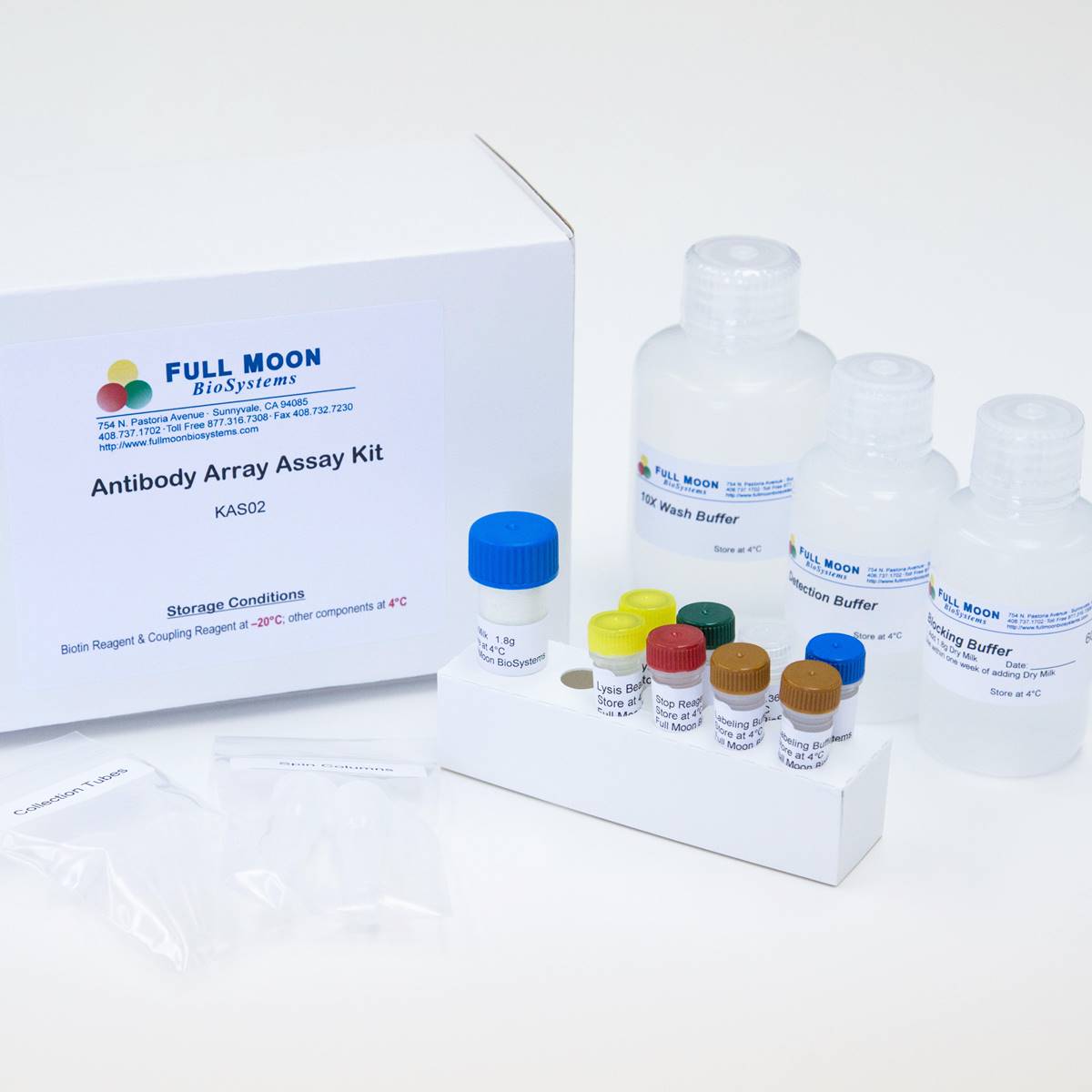
The ELISA based Cytokeleton Phospho Antibody Array platform involves four major steps:
- Protein extraction with non-denaturing lysis buffer
- Biotinylation of protein samples
- Incubation of labeled samples with antibody array
- Detection by dye conjugated streptavidin

![]() GAL File (To download, right click on the file name, then choose “Save target as”)
GAL File (To download, right click on the file name, then choose “Save target as”)
I am text block. Click edit button to change this text. Lorem ipsum dolor sit amet, consectetur adipiscing elit. Ut elit tellus, luctus nec ullamcorper mattis, pulvinar dapibus leo.
Services
If you don’t have access to a microarray, send the finished arrays to our lab for scanning. Raw scan images are delivered in tiff format.
Cost: Free
Array Image Quantification and Analysis Service includes data extraction, data organization and analysis of the array images obtained through our array scanning service.
Cost: $210 per slide
Complete Antibody Array Assay Service allows investigators to send research samples to our laboratory for analysis. There is no need to purchase the arrays and reagents and running the assays yourself. Simply select the array of your choice, and then send off the samples to our lab. This convenient hands-off approach offers quick turnaround and reliable results, saving you valuable time and resources. All assays will be performed by our highly trained scientists at our headquarter in Sunnyvale, California. Results are delivered by email in 1-3 weeks.
Cost: $1,410 per sample