WHAT WE OFFER
We simplify your protein phosphorylation research by taking care of protein profiling with Cell Signaling Phospho Antibody Array—from start to finish:
Sample Processing
Send your cell lysates or protein extracts to us, and our experienced technicians will take care of all sample preparation steps according to Full Moon BioSystems’ validated protocols.
Assay Execution
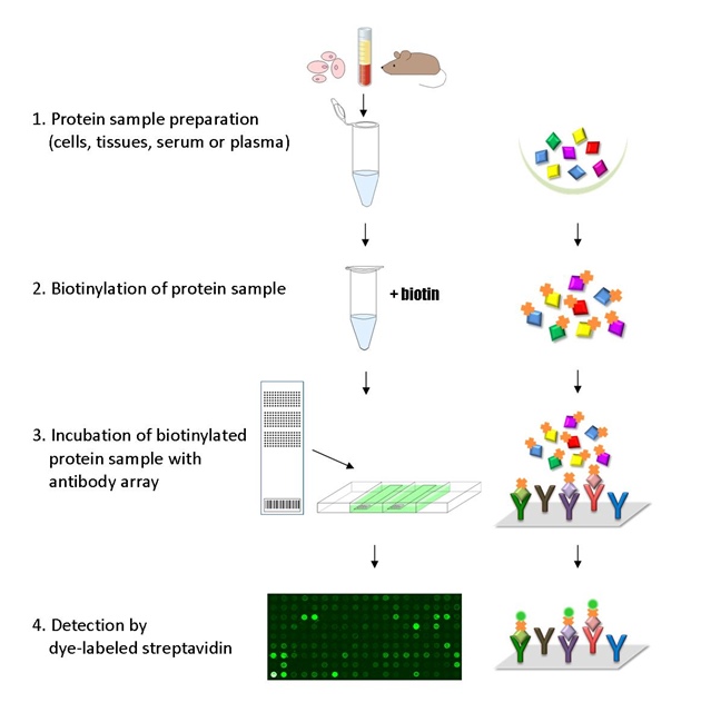
We perform the complete lysate preparation, labeling, incubation, and detection process using the Cell Signaling Phospho Antibody Array, ensuring high sensitivity and specificity for phosphorylated and total protein targets.
Imaging & Data Analysis
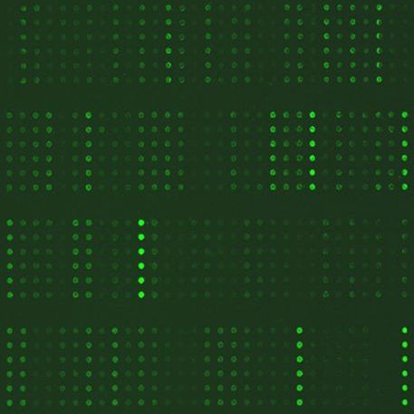
Our lab uses high-resolution microarray scanners and advance software tools to extract and analyze fluorescent signals from array images. Each spot is carefully examined to verify signal quality, consistency, and accuracy across the array.
This combined computational and expert-reviewed approach ensures high-confidence, reproducible results. We focus on delivering clean, reliable data that reflects true biological differences. This rigorous quality control process ensures that only high-confidence data are carried forward for interpretation.
Final Report Delivery
Upon completion of the assay and analysis, results are delivered in Excel format with clearly structured data. Each report includes:
-
Mean signal intensity of all replicate spots for each antibody
-
Coefficient of variation (CV) for replicate spots to assess signal consistency
-
Normalized signal intensities
-
Fold change calculations between control and. treatment samples
-
Ratios of phospho-antibody signals to their corresponding non-phospho antibody signals, providing insight into phosphorylation status relative to total protein levels
- Sample Assay Results
-
Protein phosphorylation profiling
-
Biomarker profiling
-
Cancer and drug target discovery
-
Signal pathway research
This array features 304 site-specific and phospho-specific antibodies from 16 commonly studies cell signaling pathways. Each antibody is printed with six replicates.
Antibody Reactivity: Human: 99% | Mouse: 87% | Rat: 66%
PI3K/AKT
AKT1, AKT2, BAD, BCL2, CASP9, CCND1, CCNE1, CDK2, CKDN1A, CHUK, CREB1, EGFR1, EIF4E, EIF4EBP1, ERBB2, FOXO3, GSK3B, HSP90AB1, IGF1R, IKBKB, IKBKG, IL4R, IRS1, ITGB3, JAK1, JAK2, KDR, MAP2K1, MDM2, MET, MTOR, MYC, NFKB1, NOS3, NTRK2, PDGFRB, PDK1, PPP2CA, PRKAA1, PTEN, PTK2, RAF1, RELA, RPS6KB1, STK11, SYK, TP53, TSC2, YWHAQ
AMPK
ACACA, AKT1, AKT1S1, AKT2, CCND1, CREB1, EEF2, EEF2K, EIF4EBP1, FOXO1, FOXO3, IFG1R, IRS1, MAP3K7, MTOR, PDPK1, PPARG, PPP2CA, PRKAA1, RPS6KB1, SREBF1, STK11, TSC2
Apoptosis
ACTB, ACTG1, AKT1, AKT2, BAD, BAX, BCL2, BID, CASP3, CASP8, CASP9, CHUK, EIF2S1, FADD, FOS, IKBKB, IKBKG, LMNA, MAP2K1, MAP3K5, MAPK9, NFKB1, NFKB1A, PDPK1, RAF1, RELA, TP53
Autophagy
AKT1, AKT1S1, AKT2, BAD, BCL2, BECN1, EIF2S1, IGF1R, IRS1, MAP2K1, MAP3K7, MAPK9, MTOR, PDPK1, PPP2CA, PRKAA1, PTEN, RAF1, RPS6KB1, STK11, TSC2
Cell Cycle
ABL1, CCND1, CCNE1, CDC25C, CDK2, CDKNIA, CHEK1, CHEK2, GSK3B, HDAC1, HDAC2, MDM2, MYC, PLK1, RB1, SMAD2, SMAD3, TP53, YWHAQ
ErbB
ABL1, AKT1, AKT2, BAD, BRAF, CAMK2A, CAMK2B, CAMK2D, CAMK2G, CBL, CDKN1A, CRK, EGFR, EIF4EBP1, ELK1, ERBB2, GAB1, GSK3B, MAP2K1, MAP2K4, MAPK9, MTOR, MYC, PAK1, PLCG1, PTK2, RAF1, RPS6KB1, SHC1, SRC, STAT5A, STAT5B ,
Focal Adhesion
ACTB, ACTG1, AKT1, AKT2, BAD, BCL2, BRAF, CCND1, CRK, CTNNB1, EGFR, ELK1, ERBB2, GSK3B, IGF1R, ITGB3, KDR, MAP2K1, MAKP9, MET, PAK1, PDGFRB, PDPK1, PPP1CA, PTEN, PTK2, RAF1, RASGRF1, SHC1, SRC, VASP
Insulin
ACACA, AKT1, AKT2, BAD, BRAF, CALM1, CBL, CRK, EIF4E, EIF4EBP1, ELK1, FOXO1, GSK3B, IKBKB, IRS1, MAK2K1, MAPK9, MTOR, PDPK1, PPP1CA, PRKAA1, RAF1, RPS6KB1, SHC1, SREBF1, TSC2 ,
JAK-STAT
AKT1, AKT2, BCL2, CCND1, CDKN1A, EGFR, IFNGR1, IL10RA, IL4R, JAK1, JAK2, MTOR, MYC, PDGFRB, PTPN11, RAF1, STAT1, STAT2, STAT3, STAT4, STAT5A, STAT5B, STAT6
MAPK
AKT1, AKT2, BRAF, CHUK, CRK, EGFR, ELK1, ERBB2, FOS, HSPB1, IGR1R, IKBKG, KDR, MAP2K1, MAP2K4, MAP3K5, MAP3K7, MAPK14, MAPK9, MAPT, MET, MYC, NFKB1, NTRK2, PAK1, PDFGRB, RAF1, RASGRF1, RELA, RELB, RPS6KA1, RPS6KA2, RPS6KA3, RPS6KA5, SRF, TP53, CASP3
mTOR
AKT1, AKT1S1, AKT2, BRAF, CHUK, EIF4E, EIF4EBP1, GSK3B, IFG1R, IKBKB, IRS1, MAP2K1, MTOR, PDPK1, PRKAA1, PTEN, RAF1, RPS6KA1, RPS6KA2, RPS6KA3, RPS6KB1, STK11, TSC2, WNT1
NF-kappa B
BCL2, BLNK, CHUK, IKBKB, IKBKG, LCK, LYN, MAP3K7, NFKB1, NFKB1A, PLCG1, RELA, RELB, SYK, ZAP70
p53
BAX, BCL2, BID, CASP8, CASP9, CCND1, CCNE1, CDK2, CDKNIA, CHEK1, CHEK2, MDM2, PTEN, TP53, TP73, TSC2, CASP3, CASP8
Ras
ABL1, AKT1, AKT2, BAD, CALM1, CHUK, EGFR, ELK1, GAB1, GAB2, GRIN1, IGF1R, IKBKB, IKBKG, KDR, MAP2K1, MAPK9, MET, NFKB1, PAK1, PDFGRB, PLCG1, PTPN11, RAF1, RASGRF1, RELA, SHC1, ZAP70
TGF-beta
MYC, PPP2CA, RPS6KB1, SMAD1, SMAD2, SMAD3, TGFBR1, TGFBR2
VEGF
AKT1, AKT2, BAD, CASP9, HSPB1, MAP2K1, MAPK14, NOS3, PLCG1, PTK2, RAF1, SRC, VEGFR2
Ashraf, S, Taegtmeyer H, Prolonged cardiac NR4A2 activation causes dilated cardiomyopathy in mice, Basic Res Cardiol. 2022 Jul 1;117(1):33. doi: 10.1007/s00395-022-00942-7
Chalise U, Becirovic-Agic, MMP-12 polarizes the neutrophil signalome towards an apoptotic signature, J. Proteomics. 2022. https://doi.org/10.1016/j.jprot.2022.104636
Choe YJ, Min JY, Heterotypic cell-in-cell structures between cancer and NK cells is associated with
enhanced anti-cancer drug resistance, iScience. 2002 Aug 27, 105017. https://doi.org/10.1016/j.isci.2022.105017
Jungholm O, Trkulja C, Novel druggable space in human KRAS G13D discovered using structural bioinformatics and a P-loop targeting monoclonal antibody. Sci Rep. 2024, 3;14(1):19656. doi: 10.1038/s41598-024-70217-9.
Sun X, Chen M, Knockdown of KIF15 promotes cell apoptosis by activating crosstalk of multiple pathways in ovarian cancer: bioinformatic and experimental analysis, Int J Clin Exp Pathol. 2021 Feb 1;14(2):267-291
Wang H, Ge L, 3,4,5-Tri-O-caffeoylquinic acid methyl ester isolated from Lonicera japonica Thunb. Flower buds facilitates hepatitis B virus replication in HepG2.2.15 cells, Food Chem Toxcol. 2020 March 7. doi: 10.1016/j.fct.2020.111250
Ye Y, Wang H, Polygalasaponin F treats mice with pneumonia induced by influenza virus, Inflammopharmacology. 2019 Aug 24. doi: 10.1007/s10787-019-00633-1
- Cell and tissues
- Cells: > 5 million cells
- Tissues: > 75 mg
- Lysates or protein extracts
- Protein concentration: > 2 mg/mL recommended
- Protein amount: > 400 ug
- Compatible lysis buffer: mild, non-denaturing lysis, e.g., RIPA, T-per, M-per (avoid high salt/detergent concentrations)
- Samples are stored at −80°C prior to shipping
- Assay Service Guide (Includes detailed sample preparation and submission instructions and forms)
- Samples must be frozen and shipped on dry ice in an insulated container
- Include sufficient dry ice for 48+ hours of transit time. Arrange shipment to ship early in the week (Monday–Wednesday for domestic shipments; Monday for international shipments) via overnight or international priority service.
- Email package tracking number to moc.o1749492375ibnoo1749492375mlluf1749492375@trop1749492375pus1749492375 to notify us of incoming shipments.
- Documentation
- A complete Sample Submission Form (MS WORD) placed inside your shipping container.
-
If submitting from outside the U.S., include required customs documents:
- Commercial invoice
-
Shipper’s Declaration stating “Non-hazardous biological sample for research use only”. This declaration must be completed on your institution’s letterhead.
Price: $1,730/sample
Note: The price listed is per sample per condition. Submitting both a control and a treatment sample counts as two samples.
U.S. Customers
-
Online Orders: credit card only
-
Purchase Orders (POs): We accept institutional or corporate POs. Email your PO to moc.o1749492375ibnoo1749492375mlluf1749492375@sred1749492375ro1749492375.
International Customers
-
Prepayment Required: Payment must be made in advance by credit card or wire transfer.
-
Please contact us for a formal quote, invoice, and payment instructions.
-
We’ll guide you through the sample shipping process and required documentation.


