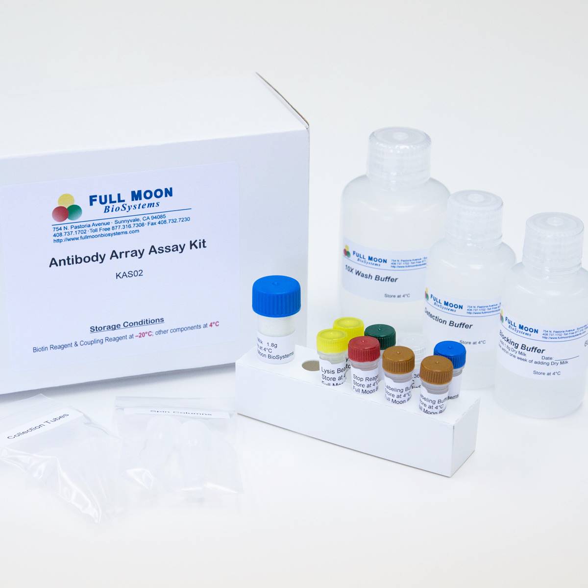
Cell Cycle Antibody Array
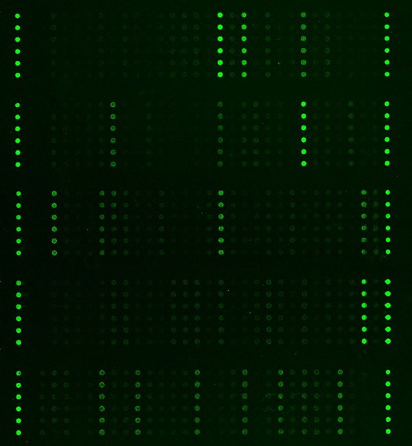
Cell Cycle Antibody Array is a high-throughput ELISA based antibody array, designed for qualitative/semi-quantitative protein expression profiling, comparing normal samples to treated or diseased samples, and identifying candidate biomarkers.
Key Features
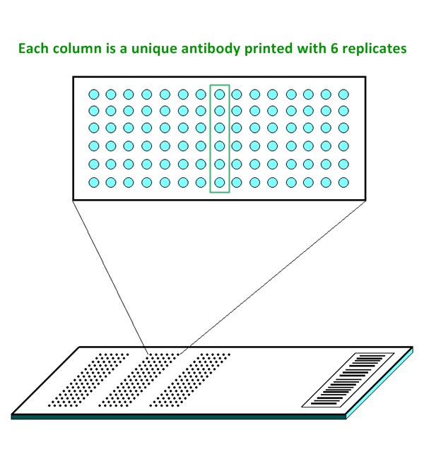
- Qualitative/semi-quantitative protein expression profiling with 62 antibodies
- Antibodies covalently immobilized on 3D polymer coated glass slide
- Fluorescent detection
Specifications
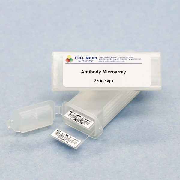
| Product Size: | 2 array slides per package for two samples (untreated vs. treated) |
| Featured Antibodies: | 62 antibodies; 6 replicates per antibody |
| Reactivity: | Human |
| Suitable Sample Type: | Cell lysate | Tissue lysate | Serum | Culture Media |
| Detection Method: | Fluorescence | Compatible Scanners |
| Internal Controls: | Positive controls: beta-actin; GAPDH | Negative controls |
| Slide dimensions: | 76 x 25 x 1 mm |
| Storage Condition: | 4°C for 6 months |
PRODUCT DETAILS
14.3.3 Pan, APC11, APC2, ATM, c-Abl, CDC14A Phosphatase, CDC6, CDC25C, CDC34, CDC37, CDC47, Cdh1, Cdk1/p34cdc2, Cdk2 , Cdk3 , Cdk4 , Cdk5 , Cdk7, Cdk8, Chk1, Cullin-1, Cullin-2, Cullin-3, Cyclin B1, Cyclin A, Cyclin C, Cyclin D1, Cyclin D2, Cyclin E, Cyclin E2, E2F-1, E2F-2, E2F-3, Glycogen Synthase Kinase 3b (GSK3b), Ki67, Mitochondria, NuMA, p130, p130cas, p14ARF, p15INK4b, p16INK4a, p18INK4c, p19ARF, p19Skp1, p21WAF1, p27Kip1, p35nck5a, p53, p57Kip2, p73, p73a, p73a/b, PCNA, RAD 51, Retinoblastoma, Retinoblastoma (Phospho-specific Ser608), ROC, Topo II beta, Tubulin-a, Tubulin-b
The ELISA based Cell Cycle Antibody Array platform involves four major steps:
- Protein extraction with non-denaturing lysis buffer
- Biotinylation of protein samples
- Incubation of labeled samples with antibody array
- Detection by dye conjugated streptavidin

![]() GAL File (To download, right click on the file name, then choose “Save target as”)
GAL File (To download, right click on the file name, then choose “Save target as”)
Basak S, Dutta S, Dengue virus modulates critical cell cycle regulatory proteins in human megakaryocyte cells. Sci Rep, 2025 15:19016
Chen P, Huang H, Bone marrow stromal cells protect acute myeloid leukemia cells from anti-CD44 therapy partly through regulating PI3K/Akt–p27Kip1 axis. Molecular Carcinogenesis 2014, doi: 10.1002/mc.22239
Hossian AKMN, Sajib MS, Multipronged activity of combinatorial miR-143 and miR-506 inhibits Lung Cancer cell cycle progression and angiogenesis in vitro, Sci Rep, 2018 8:10495
Sobol A, Galluzzo P, Depletion of Amyloid Precursor Protein (APP) Causes G0 Arrest in Non-Small Cell Lung Cancer (NSCLC) Cells,J Cell Physiol, 2015, 230(6): 1332–1341
Tian X, Jin X, The depletion of PinX1 involved in the tumorigenesis of non-small cell lung cancer promotes cell proliferation via p15/cyclin D1 pathway, Mol Cancer 2017, 16:74
Viswanathan P, Sharma Y, Replicative stress and alterations in cell cycle checkpoint controls following acetaminophen hepatotoxicity restrict liver regeneration, Cell Prolif, 2018 Mar 5. doi: 10.1111/cpr.12445
Wei X, Sun Y, Downregulation of Talin-1 expression associates with increased proliferation and migration of vascular smooth muscle cells in aortic dissection, BMC Cardiovasc Disord. 2017;17(1):162
Ye Q, Chen H, Abnormal expression of p-ATM/CHK2 in nasal extranodal NK/T cell lymphoma, nasal type, is correlated with poor prognosis, J Clin Pathol. 2021 Apr. 74(4):223-227
ADDITIONAL SERVICES
If you don’t have access to a microarray, send the finished arrays to our lab for scanning. Raw scan images are delivered in tiff format.
Cost: Free
Array Image Quantification and Analysis Service includes data extraction, data organization and analysis of the array images obtained through our array scanning service.
Cost: $160 per slide
Complete Antibody Array Assay Service allows investigators to send research samples to our laboratory for analysis. There is no need to purchase the arrays and reagents and running the assays yourself. Simply select the array of your choice, and then send off the samples to our lab. This convenient hands-off approach offers quick turnaround and reliable results, saving you valuable time and resources. All assays will be performed by our highly trained scientists at our headquarter in Sunnyvale, California. Results are delivered by email in 1-3 weeks.
Cost: $1,275 per sample