Antibody Arrays for Protein Profiling

Antibody Array for Protein Expression

Antibody Array is a high-throughput ELISA based platform for efficient protein expression profiling in cells, tissues, serum, or even culture media. Compared to the conventional methods such as Western Blots or traditional ELISA, antibody array assays are much more productive and cost-effective.

Our broad exploratory antibody arrays, including Phospho Explorer Array, Signaling Explorer Array, and Explorer Antibody Array, allow investigators to examine up to 1358 proteins on a single slide.  The pathway focused arrays are designed for researchers to study highly relevant proteins in their specific research fields.

We offer a wide range of phospho antibody arrays for protein phosphorylation profiling. The antibodies on our phospho arrays are site-specific and phospho-specific, allowing investigators to study tyrosine, serine, and threonine phosphorylation at specific sites.

Antibodies on our antibody arrays are covalently immobilized on high quality glass surface coated with proprietary 3-D polymer materials to ensure high binding efficiency and specificity. Each array includes well-characterized antibodies, positive controls, and negative controls. Each antibody is printed with two or six replicates to maximize data reliability, .

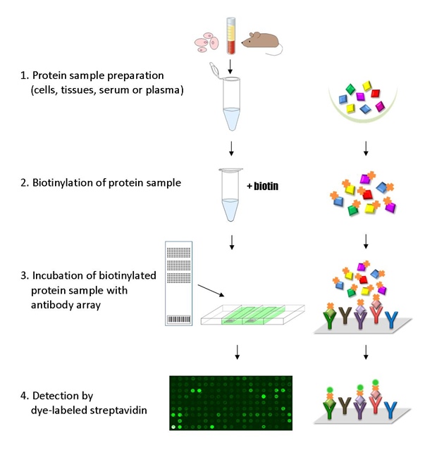
HOW ANTIBODY ARRAY WORKS

Antibody Array Assay Principle

The ELISA based antibody array platform involves four major steps:

1) Protein extraction with non-denaturing lysis buffer from cells, tissues, or serum samples

2) Biotinylation of protein sample

3) Incubation of labeled protein sample with antibody array

4) Detection by dye conjugated  streptavidin

APPLICATIONS

  • Qualitative protein expression profiling
  • Compare profiles of normal, diseased or treated samples
  • Measure phosphorylation changes at specific sites (Phospho Antibody Arrays)
  • Identify candidate biomarkers

DETECTION METHOD

  • Fluorescence
  • Instrument: microarray scanner compatible with 76 x 25 x 1 mm (3 in. x 1 in. x 1mm) slides
  • Commonly used compatible array scanners

SPECIFICATIONS

  • Suitable samples: cells, tissues, serum, culture media
  • Size: 2 Slides/Pack for processing two unique samples
  • Slide dimensions: 76 x 25 x 1 mm
  • Storage Condition : 4°C for 6 months
  • Shipping Condition: 4°C with ice packs
Exploratory Antibody Arrays

Phospho Explorer Array

Phosphorylation profiling with 1318 phospho and site-specific antibodies
More info

Signaling Explorer Array

1358 antibodies; more than 20 pathways; total protein profiling
More info

Explorer Antibody Array

Human total protein profiling with 656 antibodies
More info
Pathway Antibody Arrays
Phosphorylation Profiling
Total Protein Profiling

User's Guide

Reagent Kit Needed

Frequently Asked Questions

Each pack of arrays comes with two identical slides. Each slide can be used to run a single sample. You will need two slides (1 pack) to run a pair of samples, an untreated sample and a treatment sample.

In addition to the array slides, you will need the Antibody Array Assay Kit and dye-streptavidin that is compatible with your scanner. Please see the User’s Guide for requirements of dye-streptavidin.

Compatible samples include cells, tissues, serum, culture media, and FFPE. Generally, the recommended lysate concentration is 2 ug/uL or higher with 80 – 150 ug of protein for each array.

The antibody array platform is semi-quantitative. The data provides relative fold changes between an untreated sample and a treated sample.

A microarray scanner is needed to image the arrays. Here is a list of commonly used scanners. If you don’t access to a scanner, please consider our free array scanning service.

Certain gel imagers, such as, GE Healthcare’s Typhoon Imager and Li-Cor’s Odyssey system, may be used to image analysis. However, some of the systems may not be equipped with analysis module for microarrays. Please verify your system’s capability before using.

The software application that comes with your microarray scanner can be used for image quantification/data extraction. For example, GenePix® Pro is the acquisition and analysis software for GenePix® scanners.

To expedite spot finding, GAL files are provided free of charge and can be downloaded from here. Most modern analysis software programs are compatible with GAL files. If you don’t have access to a commercial software, please consider our image analysis service or open-source software, ImageJ, made available by NIH.

Our Complete Assay Service allows researchers to send samples to our lab in Sunnyvale, California. The assay will be conducted by our experienced scientists, and results will be delivered in electronic format by email. The turnaround time depends on the number of samples to be assayed. For 2-4 samples, the current timeline is 5-15 business days. For more details, please visit our complete assay service section.