Cytoskeleton Phospho Antibody Array
Cytoskeleton Phospho Antibody Array is a high-throughput ELISA based antibody array for qualitative protein phosphorylation profiling. It is designed for studying key proteins for cytoskeletal signaling and regulation, suitable for comparing normal samples to treated or diseased samples and identifying candidate biomarkers. This array features site-specific and phospho-specific antibodies, allowing researchers to study tyrosine and serine/threonine phosphorylation at specific sites.
Key Features
- Site-specific phosphorylation profiling and screening with 141 antibodies
- Antibodies covalently immobilized on 3D polymer coated glass slide
- Fluorescent detection
Specifications
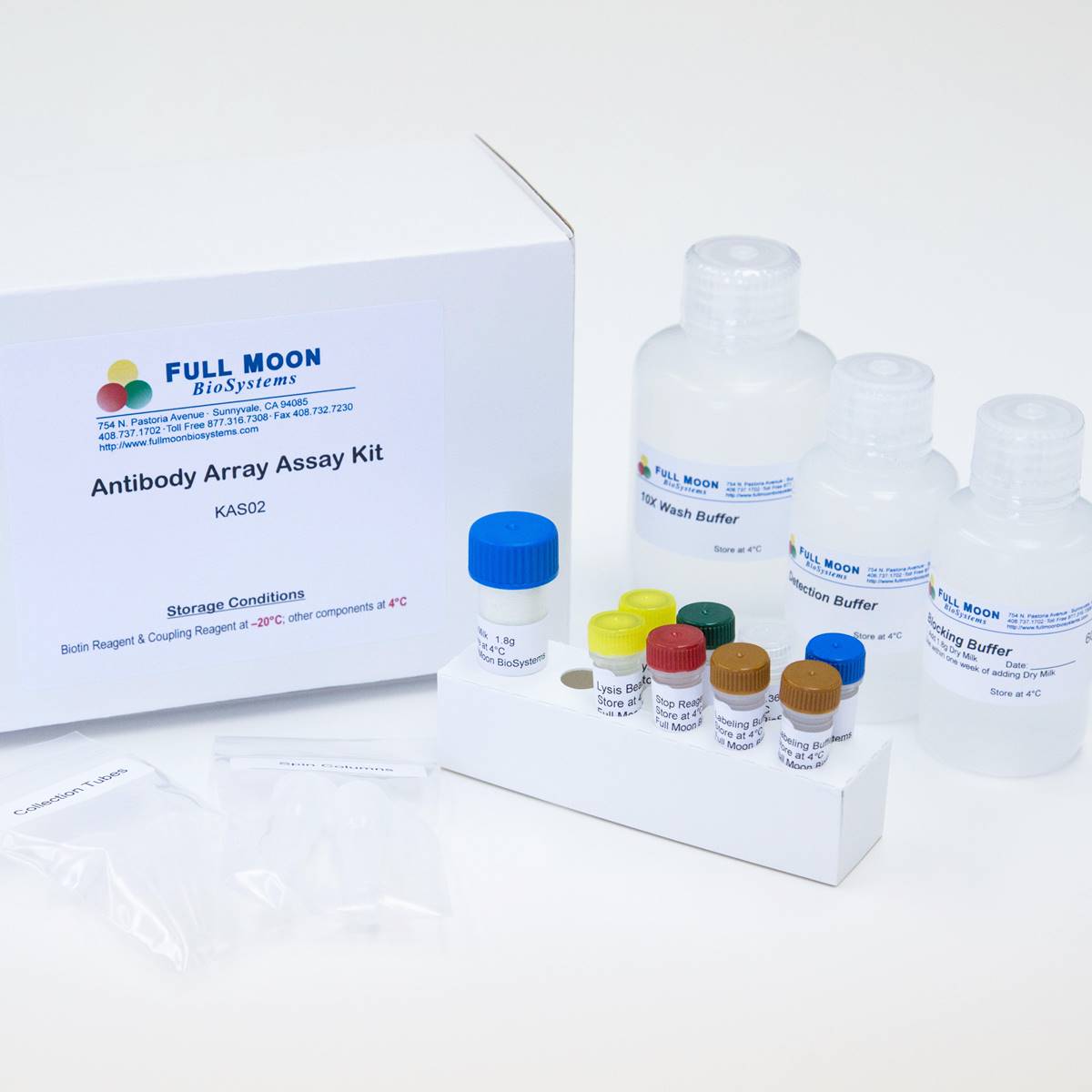
| Product Size: | 2 array slides per package for analyzing two samples (untreated vs. treated) |
| Featured Antibodies: | 141 site-specific and phospho-specific antibodies; 6 replicates per antibody |
| Reactivity: | Human: 100% | Mouse: 100% | Rat: 81% |
| Suitable Sample Type: | Cell lysate | Tissue lysate |
| Detection Method: | Fluorescence | Compatible Scanners |
| Internal Controls: | beta-actin | GAPDH | Negative controls |
| Slide Dimensions: | 76 x 25 x 1 mm |
| Storage Condition: | 4°C for 6 months |
PRODUCT DETAILS
Actin Pan (a/b/g) (Tyr55/53), Actin alpha-2/3, Actin-alpha1 sk muscle, ACTN1, Calmodulin (Thr79/Ser81), CaMK1-alpha (Thr177), CaMK1-beta, CaMK2 (Thr286), CaMK2 (Thr305), CaMK2-b/g/d (Thr287), CaMK4 (Thr196/200), CAMK5, Cofilin (Ser3), Cortactin (Tyr421), Cortactin (Tyr466), c-Raf (Ser296), c-Raf (Ser43), CrkII (Tyr221), CrkL (Tyr207), ERK1/2, ERK3 (Ser189), ERK8 (Thr175/Tyr177), Ezrin (Thr566), Ezrin (Tyr353), Ezrin (Tyr478), FAK (Ser910), FAK (Tyr397), FAK (Tyr407), FAK (Tyr576), FAK (Tyr861), FAK (Tyr925), Filamin A (Ser2152), GRB2 (Ser159), GTPase activating protein (Ser387), LIMK1 (Thr508), MEK1 (Ser217), MEK1 (Ser221), MEK1 (Thr291), MEK1 (Ser298), MEK1 (Thr286), MEK2 (Thr394), MEKKK1, MEKKK4, Merlin (Ser10), Merlin (Ser518), MKK3 (Ser189), MKK3/MAP2K3 (Thr222), MKK6 (Se207), MKK7/MAP2K7 (Ser271), MKK7/MAP2K7 (Thr275), Myosin regulatory light chain 2 (Ser18), NCK2, p130Cas (Tyr165), p130Cas (Tyr410), p44/42 MAPK (Thr202), p44/42 MAPK (Tyr204), Paxillin (Tyr118), Paxillin (Tyr31), PI3K-p85-alpha (Tyr607), PI3K-p85-a/g (Tyr467/Tyr199), PIP5K, PIP5K (Ser307), PKA CAT (Thr197), PKC alpha (Tyr657), PKC alpha/beta II (Thr638), PKC pan activation site, PLC beta (Ser1105), PLC beta3 (Ser537), Rac1/cdc42 (Ser71), Rho/Rac GEF (Ser885), Src (Ser75), Src (Tyr216), Src (Tyr418), Src (Tyr529), VASP (Ser157), VASP (Ser238), WASP (Tyr290), WAVE1 (Tyr125)
The ELISA based Cytokeleton Phospho Antibody Array platform involves four major steps:
- Protein extraction with non-denaturing lysis buffer
- Biotinylation of protein samples
- Incubation of labeled samples with antibody array
- Detection by dye conjugated streptavidin

![]() GAL File (To download, right click on the file name, then choose “Save target as”)
GAL File (To download, right click on the file name, then choose “Save target as”)
Amitrano A, Berry B, Optical Control of CD8 + T Cell Metabolism and Effector Functions, Front Immunol. 2021 Jun 3;12:666231. doi: 10.3389/fimmu.2021.666231
Boix O, Martinez M, pTINCR microprotein promotes epithelial differentiation and suppresses tumor growth through CDC42 SUMOylation and activation, Nat Commun. 2022 Nov 11;13(1):6840. doi: 10.1038/s41467-022-34529-6
Chen C, Shenoy AK, Suppression of lung cancer progression by isoliquiritigenin through its metabolite 2, 4, 2′, 4′-Tetrahydroxychalcone, J Exp Clin Cancer Res, 2018, 37(1):243
Duvall MG, Fuhlbrigge ME, Human NK Cell Cytoskeletal Dynamics and Cytotoxicity Are Regulated by LIM Kinase, J Immunol. 2020 Jul 8;ji2000186. doi: 10.4049/jimmunol.2000186
El-Haibi CP, Singh R, Antibody Microarray Analysis of Signaling Networks Regulated by Cxcl13 and Cxcr5 in Prostate Cancer. Journal of Proteomics and Bioinformatics 2012, 5(8): 177-184
Georgouli M, Herraiz C, Regional Activation of Myosin II in Cancer Cells Drives Tumor Progression via a Secretory Cross-Talk with the Immune Microenvironment, Cell. 2019 Feb 7;176(4):757-774.e23
Li F, Chen T, Hu S, Superoxide Mediates Direct Current Electric Field-Induced Directional Migration of Glioma Cells through the Activation of AKT and ERK, PLoS ONE 2013, 8(4): e61195. doi:10.1371/journal.pone.0061195
Pan Z, Dumas E, Bacillus anthracis Edema Toxin Inhibits Efferocytosis in Human Macrophages and Alters Efferocytic Receptor Signaling, Int. J. Mol. Sci. 2019, 20(5), 1167
Schubert KM, Qiu J, The AMP-Related Kinase (AMPK) Induces Ca2+-Independent Dilation of Resistance Arteries by Interfering with Actin Filament Formation, Circulation Res, 2017 121:149-161
Woollard S, Li H, HIV-1 induces cytoskeletal alterations and Rac1 activation during monocyte-blood-brain barrier interactions: modulatory role of CCR5, Retrovirology 2014, 11:20
Yamada S, Yassin M, Unique osteogenic profile of bone marrow stem cells stimulated in perfusion bioreactor is Rho-ROCK-mediated contractility dependent, Bioeng Transl Med. 2023;e10509. doi:10.1002/btm2.10500
ADDITIONAL SERVICES
If you don’t have access to a microarray, send the finished arrays to our lab for scanning. Raw scan images are delivered in tiff format.
Cost: Free
Array Image Quantification and Analysis Service includes data extraction, data organization and analysis of the array images obtained through our array scanning service.
Cost: $200 per slide
Complete Antibody Array Assay Service allows investigators to send research samples to our laboratory for analysis. There is no need to purchase the arrays and reagents and running the assays yourself. Simply select the array of your choice, and then send off the samples to our lab. This convenient hands-off approach offers quick turnaround and reliable results, saving you valuable time and resources. All assays will be performed by our highly trained scientists at our headquarter in Sunnyvale, California. Results are delivered by email in 1-3 weeks.
Cost: $1,370 per sample